The Anatomy of the Foot and Common Problems

Reviewed and approved by the nurse Leidy Mora Molina
All the organs, elements, and systems of the human body work in complete harmony. The lower limbs are essential for the mobilization, movement and support of the body. So, are you interested in finding out all about the anatomy of the foot? Don’t turn away!
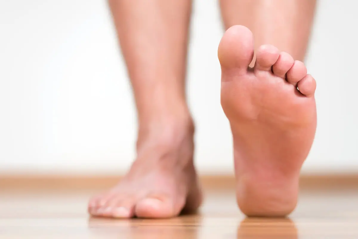
The feet are responsible for providing support, cushioning, and balance when walking, running, and practicing various physical activities that require mobilization.
It’s divided into the rearfoot, midfoot and forefoot.
Bunions, corns and warts are some of the most common foot problems. Age, excess weight and improper footwear are the main causes of these disorders. In most cases, the conditions can be solved with medical assistance.
Let’s find out what feet really are!
Feet are the end extremities of the lower limbs, responsible for supporting the entire weight of the human body and allowing locomotion. They are a complex and resistant anatomical design, and they’re the main point of contact with the environment.
This organ works in coordination and harmony with the rest of the lower limb sections to facilitate walking, running, climbing, and support when standing. Despite what we may think, even when we’re standing, there are several active muscles, ligaments, and tendons that support us.
Functions
The main function of the human foot is to serve as a base of support, and balance for the whole body. Likewise, the feet participate, together with the spine and legs, in maintaining an upright position when walking and when adopting other positions, such as sitting or standing.
In addition to this, they provide stability by adapting to various floor surfaces. Their dynamic cushioning capacity makes it possible to take advantage of the impacts when walking and to reuse the energy produced to coordinate continuous locomotion. For this reason, abnormal positions of the foot when moving can lead to different conditions, such as dislocations or fractures.
Their sensitive innervation is designed to provide information on the distribution of loads, stress and pressure in each step and position. Like the hands, they can discriminate objects, textures, and temperature changes.
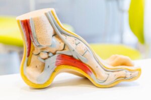
The general anatomy of the human foot
From an anatomical and functional point of view, the foot is a mechanical structure made up of 26 bones, 33 joints, 19 muscles, and more than 100 ligaments and tendons, according to experts on the subject. It’s divided topographically into 3 segments and has distinctive characteristics in the plantar area.
The forefoot, midfoot, and rearfoot are the parts that make up the anatomy of the human foot. Each segment is made up of several bones, muscles and special joints which we will describe below.
Forefoot
It’s the most distal part of the foot, also called the forefoot section. At this level, we find the metatarsals and the phalanges that form the toes.
In total, there are 5 metatarsal bones per foot and 3 phalanges for each toe, with the exception of the big toe, which only has 2 phalanges. Its main function is to give impulse and dynamics when walking.
Midfoot
This is the middle segment of the anatomy of the human foot, made up of 5 bones of medium size. They’re the cuboid bone, the navicular or scaphoid, and the 3 wedge-shaped bones or wedges, which form the plantar arch. Its basic function is to cushion and it’s susceptible to injury.
Hindfoot
This is the back section of the foot, and it contains the 2 largest bones. At this level we locate the talus and the calcaneus, which are responsible for forming the ankle and joining the foot with the rest of the leg. This segment allows the movements of the ankle and facilitates the distribution of loads.
Sole of the foot
This is the surface of the foot that makes contact with the ground. It’s made up of a thicker epidermis than the rest of the foot, especially in the support areas.
Unlike the skin in the rest of the body, the sole has an abundance of sweat glands and almost no melanocytes or sebaceous glands. Excessive sweating favors bad odors.
Similarly, this section of the anatomy of the foot has several ligaments and bony elements that form the plantar vault, supported by the medial, lateral and transverse arches.
The bones that make up the foot
The bones that make up the foot are the talus, calcaneus, navicular, cuboid, the 3 cuneiform, the 5 metatarsals, and the 14 phalanges. Each one has specific characteristics and functions:
- Calcaneus: This is the largest bone in the foot. It’s the first to receive impact when walking and provides cushioning.
- Astragalus is the second largest bone in the foot. It articulates on a lower level with the calcaneus and higher up with the tibia and fibula, forming the ankle joint.
- Navicular or scaphoid: This is a bone with a semilunar form that joins the bones of the tarsus with those of the metatarsus, offering stability. The posterior tibial tendon inserts into it.
- Cuboid: This is a cube-shaped bone located on the side of the foot. Its function is to transmit the dynamics of the ankle to the rest of the foot.
- Cuneiforms: These are 3 wedge-shaped bones, responsible for forming the tarsometatarsal joint in union with the cuboid.
- Metatarsals: These are 5 elongated bones, whose head forms the base of the toes and comes into direct contact with the ground. The first metatarsal is the largest and is the key bone during walking.
- Phalanges: The human foot has 14 phalanges, 2 in the big toe (proximal and distal) and 3 in the rest of the toes (proximal, middle and distal).
Based on the length of the toes, the foot can have 3 classic shapes: square, Greek or Egyptian.
You may be interested in reading: Caring For Your Hands and Feet
Foot muscles
The intrinsic dynamic elements of the foot are classified into dorsal muscles and plantar muscles, according to experts. The dorsal muscles are the extensor digitorum brevis and extensor digitorum brevis of the big toe. The plantar muscles are grouped together in 4 layers:
- First layer: Formed by the abductor pollicis, flexor digitorum brevis and abductor digiti minimi muscles.
- Second layer: This includes the quadratus plantaris and lumbrical muscles.
- Third layer: This is a fairly dense layer, consisting of the adductor pollicis, flexor pollicis brevis, flexor digitorum brevis of the fifth toe, and the opponens of the fifth toe.
- Fourth layer: This includes the dorsal and plantar interosseous muscles.
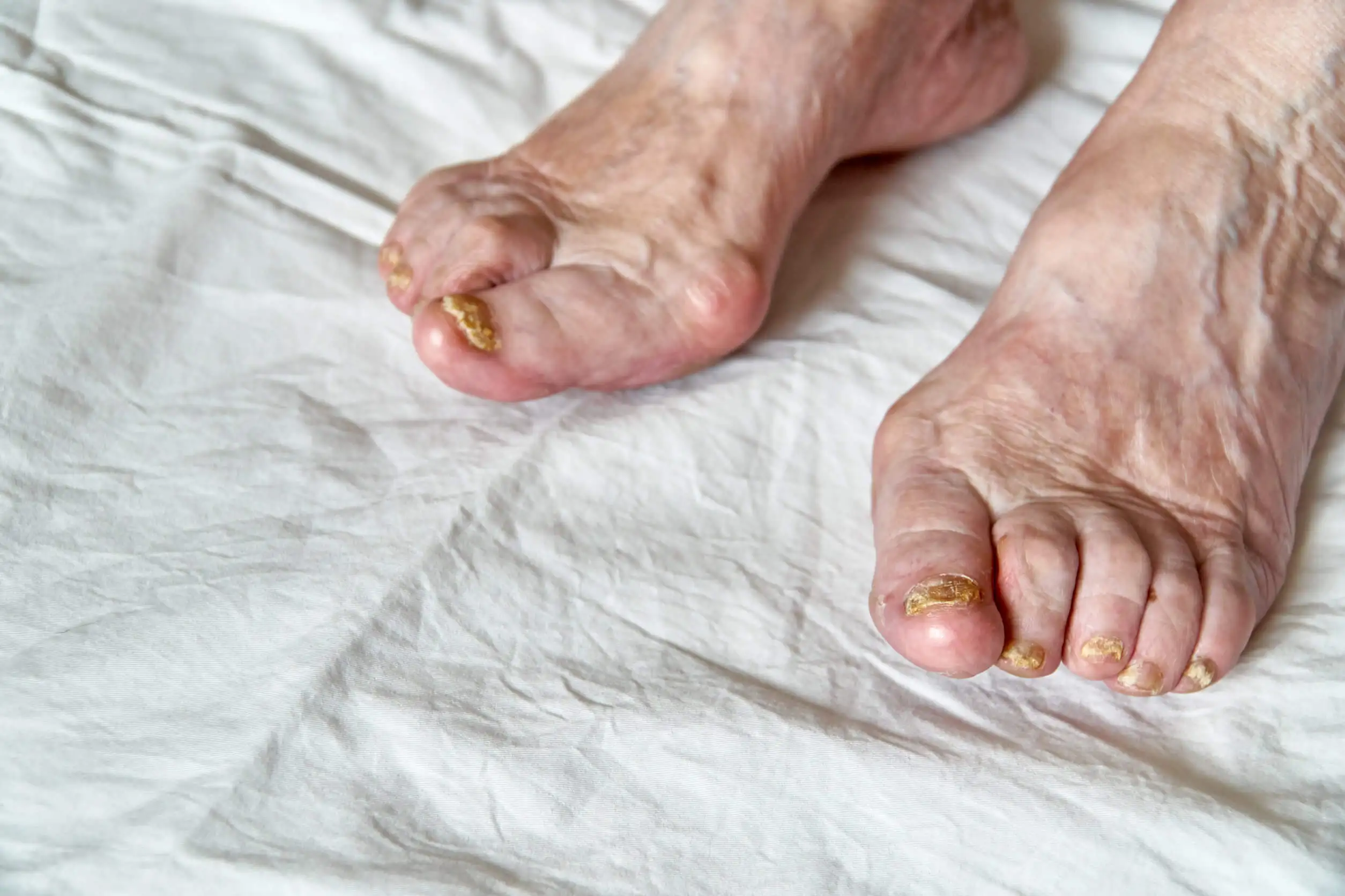
Common foot problems
Feet are prone to various conditions as a result of daily activities, improper footwear, obesity, diabetes, and aging. Some of the most common foot problems are as follows:
- Bunions: These are hard and painful protuberances in the joint of the toe.
- Calluses are patches of thickened skin, resulting from friction and pressure on the foot.
- Warts are hard, rough lesions that form on the sole of the foot and heel. Studies affirm that they’re produced by the human papillomavirus (HPV) 1, 2, 4, 571-3.
- Athlete’s foot: This is a fungal infection of the foot that thrives in dampness. It usually occurs with itching, redness, cracking and peeling.
- Ingrown toenails: This is the growth of the nail beyond the edges, penetrating the skin of the toes. This condition causes pain and great discomfort.
- Claw toes: The toes adopt a claw position because of prolonged use of tight shoes. It can also result from diseases such as diabetes, rheumatoid arthritis and brain injuries.
Foot care
To preserve foot health, we must take into account 3 factors: hygiene, footwear, and exercise. Some recommendations for efficient care are the following:
- Wash daily with lukewarm water and neutral soap
- Dry very well after bathing and avoid humidity
- Massage with moisturizing creams
- Exfoliate once a week with a pumice stone
- Cut the nails straight, without giving them a rounded shape
- Use breathable cotton socks
- Wear soft, comfortable shoes
- Avoid the prolonged use of high heels or narrow shoes
Similarly, it’s advisable to practice exercises to keep your feet healthy. Such is the case of walking on sand or grass barefoot to improve flexibility. Also, try tiptoeing, ankle rotations and picking up objects with your feet.
Find out more here: Say Goodbye Swollen Feet By Using These Homemade Remedies
A complex structure and key to mobilization
The anatomy of the foot includes a wide variety of bone, muscle and joint systems that act as levers during movement and distribute loads while standing. Changes in the foot compromise its functionality, affecting gait and balance. If in doubt, consult a specialist.
All cited sources were thoroughly reviewed by our team to ensure their quality, reliability, currency, and validity. The bibliography of this article was considered reliable and of academic or scientific accuracy.
- Fustero I. Cuidado de los pies. Offarm. 2007; 26(2): 66-72.
- Viladot A. Anatomía funcional y biomecánica del tobillo y el pie. Revista Española de Reumatología. 2003; 30(9):469-377.
- López-Giménez M. Tratamiento eficiente de 5 casos de verrugas plantares recalcitrantes con imiquimod 5%. Actas Dermo-Sifiliográficas. 2013;104(7):640-642.
- McNutt EJ, Zipfel B, DeSilva JM. The evolution of the human foot. Evol Anthropol. 2018 Sep;27(5):197-217.
- Phillips RD. The normal foot. J Am Podiatr Med Assoc. 2000 Jul-Aug;90(7):342-5.
This text is provided for informational purposes only and does not replace consultation with a professional. If in doubt, consult your specialist.


