Types of Cartilage: Characteristics and Location

Reviewed and approved by the nurse Leidy Mora Molina
There are three types of cartilage that are characterized according to their characteristics and location: hyaline, elastic, and fibrous. However, they all have some aspects in common, such as the fact that they constitute a special form of connective tissue.
Furthermore, all of the different types of cartilage are firm, flexible, and distributed in various parts of the body, from the bones to the nose, ears, and trachea, among others. Their functions are varied; for example, in the joints, they help to avoid friction between the bony parts and help resist impact.
So, do you want to know more about the different types of cartilage?
What is cartilage?
There are various types of tissue in the body. One of these is the so-called “connective tissue”, which fulfills relevant functions such as providing structure, support, and protection to other tissues and organs, as well as storing and helping in the transport of various substances.
Connective tissues include blood, lymph, adipose tissue, cartilage, and bones. The last two make up the osteoarticular system. Even though they’re related and have similar functions, they also differ in several key characteristics.
It’s interesting to note that most of the large bones of the fetus are first formed as cartilage. They’re then replaced through a process known as endochondral ossification.
Cartilage is often found in joints, although not exclusively, as it’s also present in the ears, nose, trachea, and other areas of the body.
This specialized connective tissue is made up of an extracellular matrix, for the most part (95%). It consists of various types of collagen as well as hyaluronic acid. To a lesser extent, different cells are found, such as the following:
- Fibrocytes
- Fibroblasts
- Chondroblasts
- Chondrocytes
The latter has the function of performing the synthesis of collagen fibers. Finally, there are the Proteoglycans, which are extracellular matrix molecules containing proteins and polysaccharides and are responsible for the compressibility of cartilage.
The characteristics of cartilage tissue
Cartilage has some characteristics that distinguish it from other connective tissues. Among these, the following stand out:
- Within it, there are spaces called chondroblasts or lacunae, where chondrocytes are located.
- Chondrocytes are mature cells and chondroblasts are immature cells.
- Cartilage does not have nerve endings or blood vessels.
- For this reason, it doesn’t usually have color or experience any sensitivity.
- Its cells are nourished through a process of passive diffusion by means of a matrix.
We think you may also enjoy reading this article: Weak Bones and Joints: Learn How to Strengthen Them
The functions of cartilage
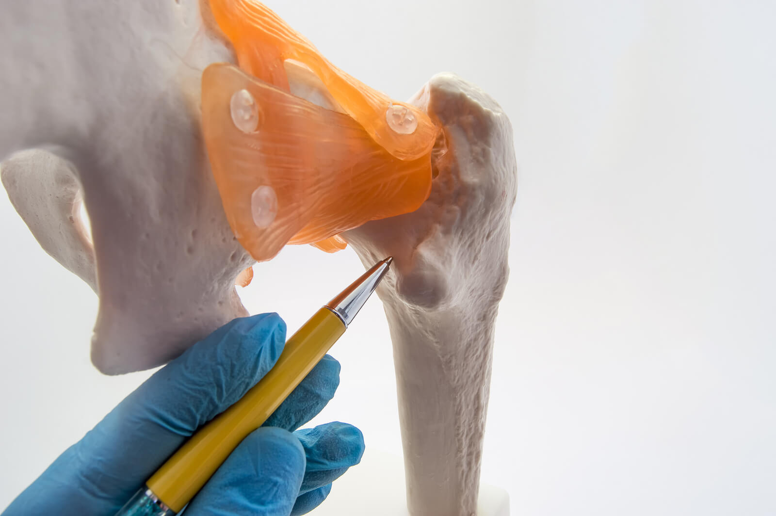
First of all, cartilage is located on the articular surfaces of the bones and, between the bones, i.e. in the joints. It thus allows mobility, while acting as a shock absorber to resist the forces exerted on the body.
Accordingly, synovial fluid is deposited on the cartilage, which acts as a lubricant to reduce friction in the joints allowing movement.
On the other hand, it can also be located between two bones, without being a joint itself. For example, this occurs in the sternum and ribs. In this case, it acts more like a means of fixation.
Finally, we also have types of cartilage such as those of the ears and the nasal septum, which serve to shape these structures, as well as the cartilage of the trachea, which constitutes a reinforcement of the trachea.
Different types of cartilage
According to their characteristics and functions, there are several different types of cartilage: hyaline, elastic, and fibrous. Let’s take a look at each of them in more detail.
1. Hyaline cartilage
Hyaline is considered the most abundant form of cartilage in the body. This type of cartilage is present in the articular ends of the larger bones and ribs, as well as in the nose, trachea, larynx, and even the bronchi.
It consists of very few fibers and is covered by a connective tissue sheath, the perichondrium. However, there’s no perichondrium in the epiphysis or head of long bones.
Its chondrocytes are organized in clusters, called “isogenic” or “isogenic” groups. Since they lack blood supply, they’re nourished by synovial fluid.
Hyaline cartilage contains several types of collagen, although type II collagen predominates in its matrix. It also contains proteoglucans and other non-collagenous proteins. It’s whitish in appearance, with pigmented areas around the lacunae, in which proteoglucans predominate.
2. Elastic cartilage
As its name implies, this type of cartilage is characterized by its flexibility. This is due to elastin and certain fibers, as well as braided sheets of elastic material. It also contains collagen type II.
It’s found in various parts of the body, such as the following:
- The epiglottis
- The larynx (cuneiform cartilage)
- The eustachian tube
- The pinna
- The walls of the ear canal
Like the type of cartilage described above, it lacks irrigation and also has a covering (perichondrium). However, unlike the former, elastic cartilage has a greater number of isogenic groups and its coloration is moderately yellowish.
3. Fibrous cartilage
Fibrous is also called “fibrocartilage”. It contains type I collagen. It’s usually avascular. However, in some cartilage of this class, there is a bit of blood supply.
Its chondrocytes are arranged in parallel rows between the collagen bundles. Meanwhile, the fibers in the matrix are denser, so it can withstand greater tensile forces.
Because of this, it functions as a cushioning device and offers resistance against compression. Thus, it moderates stretching and helps prevent possible tears.
Structurally, it’s a combination of the other two types. However, fibrous cartilage lacks perichondrium. It is present in different parts of the body, such as:
- The insertion of the tendons in the bones
- The meniscus of the knees
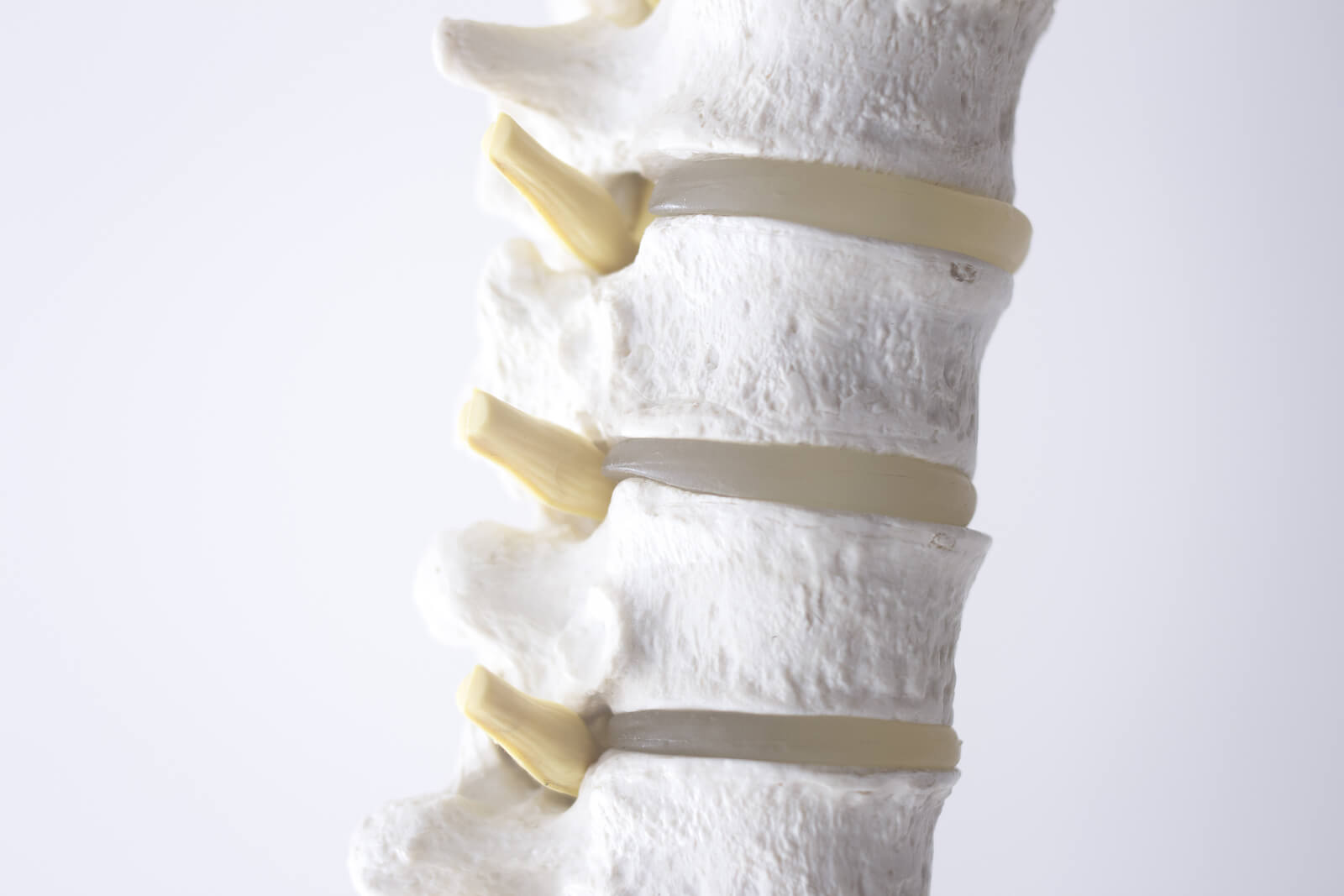
- The vertebral discs
- The sternoclavicular and temporomandibular joints
- The triangular fibrocartilaginous complex of the wrist
- The pubic symphysis
Cartilage conditions and diseases
The different types of cartilage can be affected by injury or various diseases that cause pain and inflammation, as well as deformity and limited mobility.
In sports and physical activity in general (including dance), injuries in which ligament tears and other joint damage can occur are not uncommon.
Likewise, there are diseases such as osteoarthritis, in which there is degeneration of the flexible tissue of the joint, to the point that it can cause friction between the bones.
This damage is usually a consequence of age-related degeneration. However, cases such as arthritis may be caused by an immune system reaction in which the body attacks its own tissues.
Like this article? You may also like to read: Joint Hypermobility Syndrome: What Is It and How To Deal With It?
Cartilage health
Some cartilage conditions can be prevented, although not in all cases. Avoiding sudden and repetitive movements is one of the most important measures to take care of your cartilage, especially when exercising or playing sports.
On the other hand, there are foods that help take care of these structures, such as those that are abundant in vitamin C, collagen, and omega-3. Some examples are eggs, fish, dairy products, and nuts.
Ultimately, precautions should be taken with respect to certain risk factors, such as excessive physical activity, obesity, stress, or a poor diet. These not only compromise the health of cartilage, but of all the body’s joints in general.
All cited sources were thoroughly reviewed by our team to ensure their quality, reliability, currency, and validity. The bibliography of this article was considered reliable and of academic or scientific accuracy.
- Debellea L, Tamburroa A. Elastin: molecular description and function. The International Journal of Biochemistry & Cell Biology. 1999; 31(2): 261-272.
-
Chang LR, Marston G, Martin A. Anatomy, Cartilage. [Updated 2021 Oct 21]. In: StatPearls [Internet]. Treasure Island (FL): StatPearls Publishing; 2022 Jan-. Available from: https://www.ncbi.nlm.nih.gov/books/NBK532964/
- Gorodner O. Guía Actividad 3. Sangre – tejidos cartilaginoso y óseo. Universidad Nacional del Nordeste, 2013. Disponible en: http://www.ucv.ve/uploads/media/Pr%25C3%25A1ctica_de_Cart%25C3%25ADlago_y_%25C3%25B3seo.pdf&ved=2ahUKEwjEjbK69pz7AhUzsDEKHd_6CZcQFnoECAMQAg&usg=AOvVaw3oQPNBkv3lBhXIPaT3E1uE.
- Mericq V. Factores reguladores de la osificación endocondral. Rev. Méd. Clín. Condes. 2007; 18(4): 325-329.
- Martínez-Castillo A, Núñez C, Cabiedes J. Análisis de líquido sinovial. Reumatol Clin. 2010; 6(6): 316–321.
-
Nahian A, Sapra A. Histology, Chondrocytes. [Updated 2022 Apr 21]. In: StatPearls [Internet]. Treasure Island (FL): StatPearls Publishing; 2022 Jan-. Available from: https://www.ncbi.nlm.nih.gov/books/NBK557576/
- Varios. Guías de procedimientos en reumatología. Bogotá: Asociación Colombiana de Reumatología, 2012.
- Velosa A, Teodoro W, Yoshinari N. Colágeno na cartilagem osteoartrótica. Rev Bras Reumatol. 2003; 43(3): 160-166.
This text is provided for informational purposes only and does not replace consultation with a professional. If in doubt, consult your specialist.


