What Is an Oral Fibroma and How Does it Affect Oral Health?

Written and verified by the dentist Vanesa Evangelina Buffa
The appearance of a hard lump inside the mouth is usually a cause for concern. However, this growth may be due to an oral fibroma, which is a fairly common benign growth in the mouth.
Specifically, it’s a growth of fibrous tissue that’s usually associated with situations of constant and prolonged irritation. Although it isn’t serious, it is nevertheless an alarming situation. It’s advisable to consult a physician for an accurate diagnosis. Sometimes, what appears to be a harmless lesion isn’t so harmless.
Therefore, it’s essential for the expert to evaluate its characteristics, origin, and other factors that influence its seriousness. Below, we’ll look into why they form, how they’re diagnosed and how to treat them. Continue reading!
What is an oral fibroma?
An oral fibroma is a benign tumor that appears in the soft tissues of the mouth. They can be both soft or hard lumps of scar tissue that arise in response to a constant and persistent traumatic situation.
It’s considered a reactive hyperplastic type lesion, as there’s a growth and increase in the volume of the oral mucosa’s tissues. A form of scarring develops on these tissues due to chronic irritation of the area.
It affects people of any age, although they’re more frequent in older adults. They can also appear in any area of the mouth. They’re usually small and of the same color as the oral mucosa where they’re found.
That’s why it’s sometimes difficult to notice them. However, if the factor that provokes them continues over time, then they can grow, become very evident, and even annoying.
Although oral fibroma is its best-known name, it’s also called by other names, such as the following:
- Traumatic fibroma
- Focal intraoral fibrous hyperplasia
- Oral polyp
- Fibrous nodule

Causes of oral fibromas
The most common cause of oral fibromas is chronic irritation. Trauma or constant friction on the mucosa triggers reactive hyperplasia of the tissue that gives rise to the tumor. And, if this stimulus is eliminated, the lesion disappears.
The factors and situations associated with its appearance are those mentioned below:
- The habit of biting the cheeks or lips
- The habit of sucking the lip or cheek
- Trauma to the mucosa by a broken, rough, or sharp tooth
- An irritation caused by large deposits of tartar
- Chafing caused by broken or misaligned dentures
- Injuries caused by orthodontic appliances
You should know that – less frequently – oral fibroma may have an origin other than irritation. This is the case of giant cell fibroma or fibromatosis. It tends to occur more often in older people and women. It also tends to be more common in patients with skin problems, diabetes, or anemia.
What does oral fibroma look like and how does it affect oral health?
Oral fibroma is seen in the mouth as a soft to hard, firm lump. They’re small at first, but increase in size if irritation continues. Generally speaking, they’re no larger than 1 to 1.5 cm (0.4 to 0.6 inches) in diameter.
The most common conformation is dome-shaped or as a protruding papule. However, they may have a small base stalk. In addition, if they develop under a maladaptive prosthesis, it’s common for them to be leaf-shaped.
In most cases, they’re the same color as the oral mucosa on which they’re located. However, sometimes they may be slightly paler or darker in color, especially if they have bled. After repeated trauma, their external surface may become rough, scaly, or ulcerated.
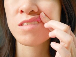
They’re most commonly located on the inner side of the cheeks, in the area where the upper and lower teeth meet. They can also be located on the inner mucosa of the lower lip, the edges of the tongue and the gums.
However, even though they’re usually painless, they sometimes cause discomfort when eating or speaking, especially when they have grown or come into contact with the teeth during chewing.
Diagnosis
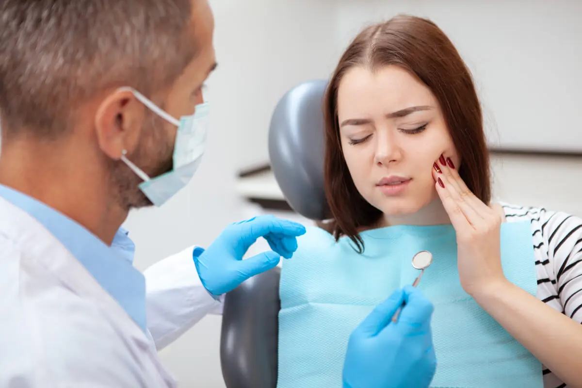
In general, when a patient has oral fibroma, it’s the patient themself who first detects it. The presence of a lump in their mouth will make them go to the dentist for a diagnosis. Even so, the lesion may also go unnoticed and it may be the dentist who discovers it in routine examinations.
Of course, the dentist is responsible for determining what it is. Intraoral examination and the presence of traumatic situations in the mouth will help to suspect an oral fibroma.
One way to confirm the diagnosis – and at the same time solve the problem – is to eliminate the irritant factor that triggers it. If the lesion decreases in size or disappears when the trauma is removed, the suspicion is confirmed.
In any case, the dentist may consider taking a sample or removing the lesion to perform a biopsy. With this histological study, an accurate and definitive diagnosis is obtained, and other similar conditions are ruled out.
If, after eliminating the irritant factor, the tumor persists, then a biopsy should be performed. This is especially the case if the patient has habits associated with oral cancer, such as alcoholism or smoking.
Differential diagnosis
Some lumps in the mouth may resemble an oral fibroma. These are some of the lesions the dentist should consider in order to make a differential diagnosis:
- Neurofibroma: This is a slow-growing mass that can be located on the tongue or buccal mucosa. However, it’s usually present in other locations as well.
- Neurilemoma: This is a tumor that’s usually located on the tongue and is accompanied by pain.
- Lipoma: A tumor similar to fibroma, but with a yellowish hue due to its adipose content.
- Peripheral ossifying fibroma: This is a focal growth located in the gum.
- Fibrosarcoma: This is a fairly rare, painless, but fast-growing malignant tumor. It usually becomes quite big and metastasizes to major organs, such as the lung.
Since the only thing that can give an accurate diagnosis is a biopsy, it’s important to perform this test and rule out other similar conditions. If necessary, the doctor will initiate appropriate treatment for the detected problem.
Treatment of oral fibroma
As we’ve mentioned, initially it’ll be necessary to eliminate the irritating factor in order to treat the oral fibroma. This may involve several measures, such as the following:
- Adapting a misaligned prosthesis
- Making a dental repair
- Polishing rough teeth
- Waxing of orthodontic wires
- Eliminating habits such as biting lips and cheeks
In any case, apart from controlling the traumatic situation, some cases warrant surgical excision of the lesion. The fibroma with narrow margins is removed and sent for biopsy to confirm the diagnosis.
You should know that, despite removing the lump with surgery, if the cause that traumatizes the tissue persists, it’s very likely that the problem will reappear. Therefore, controlling the source of the irritation is a fundamental part of the approach.
If the mucosal trauma is caused by a broken or decayed tooth, fixing it will be part of the solution. Sharp edges of fillings, dentures, or teeth should be polished or restored to avoid rubbing against the soft parts of the mouth.
For dentures that move, are loose, or that traumatize the soft tissues, it’ll be necessary to find a replacement or repair them. And if the person wears appliances that hurt, they should be refitted or covered with orthodontic wax to stop damaging the mouth.
The habit of clenching the teeth, nibbling the cheeks, or sucking on the lips can be the most difficult to control. When this habit is related to stress and anxiety, a combination of splinting, psychological counseling, and meditation and relaxation techniques can help.
If the patient has a habit of sucking mucosa through the gap left by a missing tooth, replacing the missing element is a good way to avoid this habit.
Find out more: The Dental Nerve and Toothaches: Causes and Treatments
Oral fibroid surgery
The dentist may decide that removal of the lesion is the best option to treat the problem. This is a simple procedure that the maxillofacial surgeon performs by numbing the affected area.
The fibroma is then removed with a slight safety margin using a scalpel, electroscalpel or laser. Finally, the incision site is sutured. Healing is usually quick, allowing the patient to resume daily activities as soon as possible.
The only treatment or guidelines after the treatment are:
- Maintaining oral hygiene and performing it gently in the vicinity of the treated area
- Eating a soft and cold diet for a few days
- Avoiding heat sources and exercise
- Not smoking
The tissue removed in surgery will be sent to the pathologist for a precise report on the type of lesion. Subsequent monitoring to assess the evolution of the case and the recovery of the tissues will be necessary. At this stage, the dentist will check for possible recurrences of the lesions or trauma that caused them to appear.
Preventing the formation of an oral fibroma
The best way to prevent the appearance of an oral fibroma – or to prevent it from reappearing if you have already had one removed – is to monitor your mouth carefully. Being attentive to the conditions of the oral cavity will allow you to limit the situations that can injure the mucous membranes and favor the appearance of these lumps.
Taking care of your oral health with proper hygiene and visiting the dentist regularly are two fundamental factors to prevent them. It’s also key to avoid harmful habits such as chewing on your cheeks.
If necessary, regular check-ups with the dentist will help detect lesions of greater concern in a timely manner. That said, it’s important to see a dentist when growths increase in size or are persistent.
All cited sources were thoroughly reviewed by our team to ensure their quality, reliability, currency, and validity. The bibliography of this article was considered reliable and of academic or scientific accuracy.
- Rocafuerte-Acurio, M. A. (2019). Fibroma traumático en cavidad oral–una revisión. Revista KIRU, 16(1).
- Mauri-Obradors, E., Estrugo-Devesa, A., Jané-Salas, E., Viñas, M., & López-López, J. (2017). Oral manifestations of Diabetes Mellitus. A systematic review. Medicina oral, patologia oral y cirugia bucal, 22(5), e586.
- Gnecco-Goenaga, B., Viola-Rhenals, M., & López-Arrieta, Z. (2014). Fibroma osificante periférico. Revista Ciencias Biomédicas, 5(1), 130-133.
- Gonzales, C. J. P., Abreu, J. A., & Chong, J. C. P. (2020). Fibrosarcoma del adulto. Revista Cubana de Otorrinolaringología y Cirugía de Cabeza y Cuello, 4(3).
- Centeno, A., Danielo, C., Campana, R., & Orozco, M. A. (2010). Tumores malignos de boca. Med Cutan Iber Lat Am, 38(6), 221-228.
- Sánchez Solórzano, G. A. (2019). Resección quirúrgica de fibroma traumático en la cavidad oral (Bachelor’s thesis, Universidad de Guayaquil. Facultad Piloto de Odontología).
- Dutra, K. L., Longo, L., Grando, L. J., & Rivero, E. R. C. (2019). Incidence of reactive hyperplastic lesions in the oral cavity: a 10 year retrospective study in Santa Catarina, Brazil. Brazilian journal of otorhinolaryngology, 85, 399-407.
- Zambrano, T. B. S., Mendoza, N. B., & Bazurto, M. R. N. (2020). Fibroma reactivo lateral de lengua: Presentación de un caso clínico. Revista Científica Especialidades Odontológicas UG, 3(2).
- Cobos, M. R. (2015). El fibroma traumático como lesión hiperplásica común de la boca: reporte de un caso. Ciencia y Salud virtual, 7(1), 81-87.
This text is provided for informational purposes only and does not replace consultation with a professional. If in doubt, consult your specialist.


