Black Spots on Your Gums: Causes and Treatments

Written and verified by the dentist Vanesa Evangelina Buffa
You may get a shock if, when brushing your teeth, you notice that you have black spots on your gums. These dark areas don’t only affect the aesthetics of your smile, but may also indicate that something is wrong with your oral health.
The gum tissue that lines your teeth is usually pale pink. But, just like skin, hair and teeth, each person has his or her own coloring.
The presence of black spots on the gums can be due to different causes. And, most of the time, they’re harmless situations. However, to be sure that they’re not harmful, it’s best to consult your dentist and evaluate the problem.
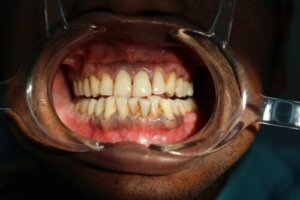
What do normal gums look like?
Gums are a soft, fibrous tissue that surrounds the main body of the teeth. They protect and cover the alveolar bone in the root area of the teeth.
Together with the bone tissue and the periodontal ligament they make up the periodontium. This means that it’s part of the structure responsible for holding and maintaining the teeth in place.
When the gums are healthy they’re pale pink, uniform, and with a dotted texture similar to orange peels. They’re firm and their defined edges surround the neck of the teeth.
But, as we already mentioned, this coloration can vary from one person to another. Just like the skin, this tissue has melanocytes, which are the cells responsible for the coloration of the epithelium.
Sometimes, the coloration of the gingival tissue changes, and you’ll need to attend to the situation. These changes in the appearance of the gums may indicate that there’s an oral health problem that should be resolved.
Contusions
Black spots on the gums may appear as a result of having suffered a trauma in the area. Like any other part of the body, gum tissue can be injured.
Banging your mouth, falling on your face, eating something with sharp edges, or even brushing your teeth too hard, can all cause bruising of the gums. The bruises appear as dark red or purple in color, but over time turn dark brown or black.
If the black spots on the gums are due to a bruise, there may also be mild pain in the area as well as bleeding.
Bruising usually resolves on its own within a few days without the need for any medical treatment. However, if the bruise persists over time and doesn’t disappear, it’s advisable to consult a dentist.
Also, if many bruises appear in different parts of the body without an apparent cause, it’s also advisable to consult a dentist. They may be a symptom of a blood problem.
Eruption cyst
When a tooth is about to erupt, a fluid-filled cyst may form on the gum. This happens because the dental crown fails to break through the gingival mucosa and come out properly.
Sometimes the accumulated fluid has blood mixed in which can give a purple or black color to the gum tissue. When an eruption cyst has blood in it, it’s called an eruption hematoma. This usually happens if the cyst has been injured by a blow or fall.
This type of process is common in childhood, during the eruption of baby teeth and permanent teeth. They disappear by themselves once the tooth comes through.
However, if this process takes longer than normal and the tooth can’t erupt on its own, then the pediatric dentist can make a small incision on the gum, like an eyelet, to facilitate the process.
Melanoplakia and melanotic macules
As we told you when talking about the appearance of normal gum, each person has their own gum tissue color. And heredity and genetics have a lot to do with the appearance of this mucosa.
This is why some patients have dark brown or black gums, and it’s due to an increased concentration of melanin on their gingival tissue. This situation is called melanoplakia and is considered harmless and normal.
These pigmentations can occur in any area of the gingiva and are usually on both sides. They’re asymptomatic and become more noticeable as a person gets older. They’re also commonly seen on the mucosa of the cheeks and lips.
They’re more common in certain ethnic groups and were formerly called racial spots or racial melanic pigmentation.
Melanotic macules, on the other hand, are harmless spots similar to freckles. They can occur on several parts of the body, including the gums.
Melanotic macules are solitary brown, bluish, or black spots that are asymptomatic. They’re small, usually between 1 and 8 millimeters in diameter, and well circumscribed.
The cause of these lesions is unknown and some people are born with them. No treatment is necessary. However, if a more serious pathology is suspected, the dentist may suggest a biopsy.
Smoking
Among the habits that are harmful to oral health, smoking is one of the most damaging. This habit doesn’t only stain teeth. Heat and toxins damage the oral mucosa and can cause serious pathologies.
People who have been smoking for several years often have black spots on their gums. Nicotine increases the production of melanin in the cells, giving rise to dark gingival tissue. This is known as smoker’s melanosis.
A darkening of the mucous membranes can also be seen on the lips and cheeks. There’s a link between the decrease in this dark pigmentation and smoking cessation. Therefore, black spots on the gums are considered reversible.
Read more here: 5 Ways Quitting Smoking Improves Your Appearance
Amalgam tattoos
Black stains on the gum due to amalgam occur after the patient has had a filling made with this material. Some particulate from the filling may become lodged around the teeth.
Silver amalgam is a material used to fill cavities in teeth. If it reaches the inside of the mucosa, it’s capable of impregnating and coloring it. This can happen when the filling is made or when it’s removed with rotary instruments to replace it with resins.
The stains are usually located near the fixed tooth. They’re dark blue or black in color.
They’re asymptomatic and permanent, and they aren’t removed, as it isn’t necessary. But, even though they aren’t serious, they can still be an aesthetic nuisance if they’re very visible.
To prevent this type of black spot on the gums, the dentist should use dental dam if they’re going to place or remove amalgams. In this way, the soft tissues are isolated.
The use of certain medications
The intake of certain medications can cause, as a side effect, the darkening of the gums. Oral contraceptives of the combined type are the most common example.
Some antibiotics, antimalarials, antipsychotics, and anticancer drugs can also cause black spots on the gums. Among these, chlorpromazine, minocycline, and quinidine have presented this type of undesired effect.
Periodontal diseases
Gingivitis and periodontitis can also give rise to black spots on the gums. This isn’t the most common situation, as most of the time the gum tissue becomes inflamed and looks red and bleeding.
But, when a lot of bacterial plaque accumulates and calcifies, it turns into calculus. This calculus can look dark in color and, if deposited in the subgingival area, can give the tissue a black appearance.
It’s also possible for the gums to look black temporarily after having dental treatment in the area. After a gum graft is placed to treat gingival resection, for example, the tissue will appear black for a few days because of transient necrosis that will disappear as the area heals.
Another periodontal problem that can cause black spots on the gums is acute necrotizing ulcerative gingivitis. This is an uncommon infection of the gum tissue that is also known as trench mouth.
It causes fever, bad breath, and a lot of pain in the gums, whose papillae become necrotic and appear gray or black. It’s usually the result of having suffered from gingivitis previously, in which bacteria proliferate very fast and the tissue dies. It’s associated with poor oral hygiene, an unhealthy diet, stress, lack of sleep, or immune problems.
Its treatment consists of periodontal cleaning and the use of antibiotics. Oral hygiene and the use of mouth rinses also promote tissue recovery and prevent a recurrence.
Blue nevus
A blue nevus is a round, flat or slightly elevated black or bluish mole located on the gum (and other parts of the skin). It’s like a freckle on the gingival tissue.
The etiology is unclear and it usually develops during childhood or adolescence. They’re more frequent in women.
In general, they don’t require treatment, although it’s important to monitor them. If their size, shape, or color changes, the doctor will recommend a biopsy to detect a possible malignant transformation.
Melanoacanthoma buccal
Melanoacanthoma buccalis is a rare disorder that causes the appearance of dark spots in different parts of the mouth, including the gums. They’re harmless and usually appear in young people.
Their origin is unknown, although they seem to be related to injuries caused by chewing or friction in the mouth. They don’t require any treatment, although the similarity with other lesions merits a biopsy to rule out more serious pathologies.
Oral cancer
Oral cancer can also cause black spots on the gums. There are other symptoms associated with this pathology, such as open sores that don’t heal, unusual bleeding, swelling, a chronic sore throat, or voice changes. Although the stain can also be the only manifestation.
To determine whether a black spot is caused by cancer, the physician must perform a biopsy. This can be complemented by different imaging techniques, such as a CT scan or PET scan. Especially to see if the cancer has spread to other parts of the body.
If the spot is malignant and the cancer hasn’t spread, the doctor may remove it surgically. If it has spread, chemotherapy and radiation therapy will help kill the cancer cells.
Patients who smoke and consume large amounts of alcohol are at increased risk of developing oral cancer.

Find out more here: Risk Factors for Mouth Cancer
Other pathologies
Some systemic conditions can also be responsible for the appearance of black spots on the gums. Determining the presence of these pathologies is essential in order to decide on appropriate treatment to address the cause of the problem.
One example is Peutz-Jeghers syndrome. This is an autosomal dominant inherited condition that causes oral and perioral melanic pigmentations. It also increases the risk of developing intestinal polyps and cancer. A genetic test is necessary in this case.
Addison’s disease can also give rise to black spots on the gums and other oral mucosa. It’s an autoimmune disease that affects the adrenal glands.
Addison’s disease patients often have fatigue, extreme thirst, weight loss, a lack of appetite, and muscle weakness. In addition, there’ll be hyperpigmentation of the lips, palate, gums, and dark patches in other areas of the body.
Treatment for black spots on gums
To treat black spots on gums it’s necessary to find out their origin and act accordingly. However, there are some methods for cosmetic purposes that can improve the appearance of gum tissue when the darkening is benign.
Here are some possibilities to improve the appearance of dark gums:
- Microabrasion: This is the removal of the superficial layers of the gum with specific instruments. This discards the pigmented area and exposes the healthy, pale pink tissue underneath.
- Surgical removal: This consists of removing the stains from the gums by cutting and removing them with a scalpel. This is an invasive technique with a more complicated postoperative period. There’s a healing process for the tissue and the results aren’t always long-lasting.
- Cryosurgery: This is a procedure in which the affected gingival tissue is frozen.
- Free gingival grafts: This treatment consists of removing non-pigmented tissue from the palate and placing it in the gum area to hide the pigmentation.
- Laser: This is the elimination of melanin-producing cells using laser technology. It’s a minimally invasive procedure, which reduces bleeding, and favors the patient’s prompt recovery.
Many of these practices can injure the mucosa or the stains may reappear. It’s important to clarify all these aspects with your dentist before starting treatment.
All cited sources were thoroughly reviewed by our team to ensure their quality, reliability, currency, and validity. The bibliography of this article was considered reliable and of academic or scientific accuracy.
- Flores, A. G. F., & Oviedo, D. M. G. (2018). Quiste de erupción: Reporte de caso. Revista Mexicana de Estomatología, 5(1), 55-56.
- de la Teja Ángeles, E., Garza-Elizondo, R., & Durán-Gutiérrez, L. A. (2021). Erupción dental difícil. Acta Pediátrica de México, 42(4).
- Blanco Carrión, A., Somoza Martín, M., & López López, J. (2003). Discromías de la mucosa oral. Gaceta Dental, 2003, num. 136, p. 32-50.
- Calderón García, P. A. (2021). Gingivitis Ulcero necrosante y su relación con el VIH-sida (Bachelor’s thesis, Universidad de Guayaquil. Facultad Piloto de Odontología).
- Bruno, G., Zavala, M. C., Sapag, M., Arzac, E., Zanelli, M. R., Vera Tapia Brook, M. J., & Lancon, C. A. (2020). Gingivitis ulcero necrotizante aguda. In IV Jornadas de Actualización en Prácticas Odontológicas Integradas PPS-SEPOI (La Plata, 7 de julio de 2020).
- Blanco, G. F. LESIONES PIGMENTARIAS DE LA BOCA (Doctoral dissertation, Universidad Autónoma del Paraguay).
- Contreras, E., & Carlos, R. (2005). Melanoacantosis oral (melanoacantoma): reporte de un caso y revision de literatura. Medicina Oral, Patología Oral y Cirugía Bucal (Ed. impresa), 10(1), 09-12.
- Valerdi, M. L., Resendiz, J., Labastida, S., Gallegos, F., & Kimura, T. (2018). Melanoma primario en mucosa de cavidad bucal. Oral, 18(58), 1526-1529.
- Álvares, H. N. M., & Cueto, N. F. T. Melanoma oral primario. Revisión de la literatura.
- Sacoto Cantos, C. R. (2020). Efectividad de la microabrasion en el tratamiento de melanosis gingival (Bachelor’s thesis, Universidad de Guayaquil. Facultad Piloto de Odontología).
- Tello, P. M. C., Ayala, L. D. O., & Endara, A. S. C. (2020). Melanosis Gingival: diagnóstico y terapéutica de su implicación estética. Revisión de la literatura.
This text is provided for informational purposes only and does not replace consultation with a professional. If in doubt, consult your specialist.


