What Are Sacral Nerves?

Reviewed and approved by the doctor Mariel Mendoza
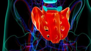
The sacral nerves are a group of five spinal nerves that come out of the spinal cord and emerge from the sacral bone. They are a set of structures that constitute the lowest segment of the spinal cord.
They are listed according to each vertebra that they relate to, although they are fused together to form the sacrum. They’re divided into specific territories, both sensory and motor types.
The sacral nerves
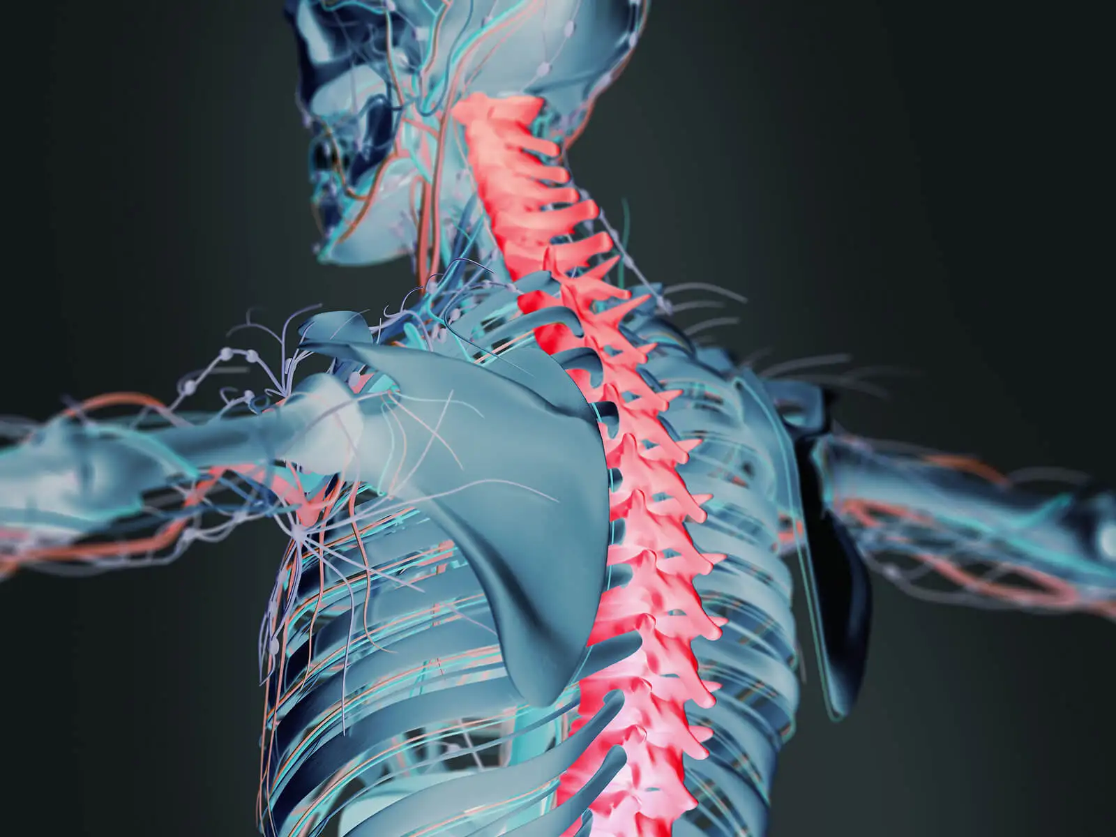
The sacral nerves are 5 pairs of spinal nerves. They form part of the somatic nervous system. More specifically, there are a total of 31 pairs of spinal nerves whose function is to innervate the entire body except for the head and some areas of the neck.
They achieve their purpose because they contain a sensory root, which is responsible for the sensitivity of the area they innervate, and a motor root, which allows the muscles to contract. The spinal nerves are as follows:
- 8 pairs of cervical nerves (C1-C8)
- 12 pairs of thoracic nerves (T1-T12)
- 5 pairs of sacral nerves (S1-S5)
- 5 pairs of lumbar nerves (L1-L5)
- 1 pair of coccygeal nerves
Read more here: 8 Tips to Keep Your Spine Healthy and Strong
Characteristics of the sacral nerves
The sacral nerves have a series of characteristics that are typical of the spinal nerves. Let us see what the main characteristics of these nerves are. To begin with, they’re mixed, i.e., both sensory and motor.
On the other hand, all the branches of the ventral branches, except the thoracic branches (T1 to T12), form several branches known as nerve plexuses. These plexuses appear in the cervical, brachial and lumbosacral areas.
Within these interconnected branches, the fibers originating from the ventral branches are cross-linked and re-distributed so that each resulting branch contains fibers from different spinal nerves. In addition, those originating from each ventral branch travel to the periphery of the body via different routes in the branches.
For this reason, all our extremities’ muscles will receive innervation from more than one spinal nerve. As a consequence, if there’s damage to one of the spinal cord segments or one of the roots, the limb need not be totally disabled.
Anatomy of the sacral nerves
The sacral nerves originate in the spinal cord and pass through the vertebral orifices of the sacrum. Both motor and sensory roots converge outside the spinal ganglion and emerge together in two branches:
- Posterior: These come out of the posterior sacral foramen and form the posterior branches of the sacral nerves.
- Anterior: These come out of the anterior sacral foramen and contribute branches to the sacral plexus and sacrococcygeal plexus.
Find out more: Bionic Spine: Helps Paralyzed Walk Again
As for the S3 and S4 sacral nerves, they have fibers that join the inferior hypogastric plexus, which innervates the smooth musculature of:
- The descending and sigmoid colon
- The rectum
- The genital organs, both internal and external
- The bladder
- The urethra
Posterior branch of the sacral nerves
These are 4 small nerve divisions that become even smaller as they become more distant from their point of origin. They emerge, with the exception of the last innermost branch, from the posterior sacral foramina.
Anterior branch of the sacral nerves
As for the anterior branches of these nerves, they are formed in the sacral plexus. This provides innervation to the pelvis and lower limbs. The bundles and fibers coming from the anterior sacral nerves (S4 and S5) join the coccygeal nerve and form, as we have seen, the sacrococcygeal plexus.
Finally, it ends up forming the anococcygeal nerve, which innervates the skin of the anal region and the sacrococcygeal joint.
Functions of the sacral nerves

The function of the parasympathetic system consists in relaxing the sphincters and contracting the muscular walls. In doing so, they cause urination, defecation, and the erection of the genitals.
In addition, the fibers carry sensory signals, such as pain and the sensation of fullness, from both the bladder and the rectum. As a curiosity, sacral nerve stimulation is one of the medical procedures used for the treatment of fecal incontinence.
In short, these are structures of vital importance to the body, like all the others in the nervous system. Getting to know human anatomy helps us to understand our health from a different perspective and to look after it correctly.
All cited sources were thoroughly reviewed by our team to ensure their quality, reliability, currency, and validity. The bibliography of this article was considered reliable and of academic or scientific accuracy.
-
Dydyk AM, Hameed S. Lumbosacral Plexopathy. In: StatPearls [Internet]. Treasure Island (FL): StatPearls Publishing; 2022 Jan.Kaiser JT, Lugo-Pico JG. Neuroanatomy, Spinal Nerves. In: StatPearls [Internet]. Treasure Island (FL): StatPearls Publishing; 2022 Jan.Khan YS, Lui F. Neuroanatomy, Spinal Cord. In: StatPearls [Internet]. Treasure Island (FL): StatPearls Publishing; 2022 Jan.
This text is provided for informational purposes only and does not replace consultation with a professional. If in doubt, consult your specialist.


