A Spot on the Lung: Main Causes and Treatment


Written and verified by the doctor Maryel Alvarado Nieto
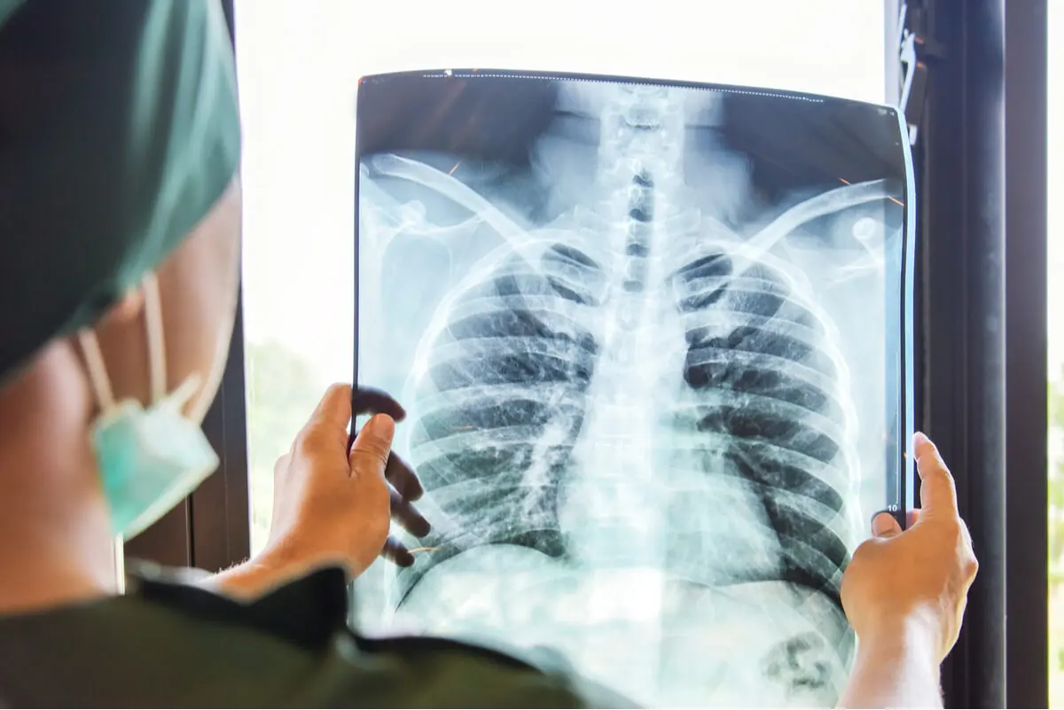
A chest X-ray is the most commonly used imaging technique in lung diseases. This study is very useful for confirming various diagnoses of the respiratory system. And this confirmation is possible thanks to the visualization of different patterns, which are actually the expression of the changes that are occurring. Among these is a spot on the lung.
The structures in the thorax have different densities, which through the incidence of x-rays, creates images that are characteristic of these densities. This constitutes a normal chest x-ray.
In popular terminology, the visualization of a whitish figure is referred to as a spot on the lung, whereas the technical term is a pulmonary nodule. However, the clinical term used will depend on the characteristics exhibited by the opacity, which is the result of increased density due to a particular pathology.
The most common causes of a spot on the lung
Infectious processes are one of the main reasons for increased density on the chest x-ray. The location and characteristics of a spot on the lung should always correlate with the patient’s clinical manifestations.
However, sometimes imaging findings may suggest a specific pathogen, allowing effective treatment to be instituted in a timely manner.
Pneumonia and bronchopneumonia
Infection of the lung tissue constitutes pneumonia, but if the ducts (bronchi) that allow air to enter the lungs are also affected, it’s known as bronchopneumonia.
The causes of this lower respiratory tract infection are viruses, bacteria, and even fungi. This spot in the lung is poorly demarcated, forming an image known as a focus of consolidation.
In the presence of a characteristic diagnosis of pneumonia or bronchopneumonia, in which the x-ray confirms the clinical findings, a course of antibiotics should be prescribed. The choice of drug will depend on the patient’s age, geographical location, and the suspected illness.
The routes of administration are related to the conditions of the patients. Generally, the intravenous route is chosen during the first days.
Pneumonia is usually treated with intravenous drugs at the beginning, and then oral therapy.Pulmonary tuberculosis
An ancient illness that’s far from being eradicated from the world is tuberculosis. Clinical manifestations include the following:
- Night sweats
- Weight loss
- Coughing up blood
A spot on the lung is evident on an x-ray. Although the image of consolidation can appear in any area of the thorax, the pulmonary apices are a frequent location.
The diagnostic suspicion must be confirmed with bacteriological studies and a sputum culture. Due to the clinical manifestations, it’s necessary to rule out any neoplastic process.
The treatment of tuberculosis consists of a course of drugs of prolonged use, so the follow-up of the cases is essential in order to be able to cure the disease.
Read more here: Foods Against Lung Inflammation
Lung abscess
The accumulation of purulent substances in a cavity formed in the lungs means it’s a lung abscess, which is a serious complication of an infectious process. The causative microorganisms lead to tissue destruction resulting in necrosis.
Clinical manifestations include the following:
- Fever
- Chest pain
- Loss of weight
- Cough with purulent and foul-smelling expectoration
Diagnostic suspicion should correlate with various risk factors, such as age, alcoholism, smoking, and secretions from the oral cavity. In the chest x-ray, it’s possible to observe a spot in the lungs formed by a whitish image, in whose interior a dark pattern can be seen.
Treatment requires hospital admission for the administration of intravenous antibiotics. Occasionally, drainage of the abscess is necessary.
Pathologies of the mediastinum: other possible causes of a spot on the lung
A chest x-ray isn’t only useful for assessing respiratory diseases. Between the two lungs, there’s a very important area: the mediastinum.
This area contains all the structures of the thorax, with the exception of the lungs. Therefore, any involvement of any of these elements will lead to the formation of a distorted image on the chest x-ray.
Tumors and masses in the mediastinum
The loss of the normal characteristics of the structures that overlap between the two lungs on the chest x-ray makes it necessary to carefully evaluate the mediastinum. These changes are sometimes a casual finding, as the illness that has caused them may not produce any symptoms.
However, the study of the radiographic findings is essential, because they may indicate a serious process.
A spot on the lung in the mediastinal space may represent both a trivial condition and be the early evidence of an extrapulmonary neoplasm. Usually, the increased density occurs on only one side of the chest, preventing the structures from being seen or displacing them on the x-ray.
In the presence of an abnormal image, a new image is requested with the patient positioned sideways (lateral projection).
In this way, the spot on the lung can be observed from two perspectives. The great usefulness of these studies lies in the fact that the mediastinum is divided into anatomical compartments, in which the different structures are constrained.
Therefore, the images can serve as a guide to locate the location of the tumor mass. Even so, a tomography is the ideal study to pinpoint both the location and the affected tissue.
Alterations of the blood vessels
In addition to masses and tumors, another cause of a spot on the lung is the existence of blood vessels that have a different shape to normal. Arteriovenous malformations are one of these alterations, which often go unnoticed because they don’t produce symptoms and their frequency is low.
However, in the presence of an anomalous vascular pattern, it’s necessary to carefully evaluate the patient. Confirmation of structural changes in the blood vessels is carried out by contrast media studies, such as angioresonance. Treatment includes arteriography with embolization and, in particular cases, surgery.
Read more: What Exactly Is a Lung Nodule?
Lung tumors are always considered
Intrapulmonary tumors also create whitish shadows on the chest X-ray. Lung cancer is one of the leading causes of oncologic death in several countries.
So a spot on the lung, without apparent cause, should be investigated taking into account the patient’s risk factors, such as age and smoking habits.
Early suspicion results in reduced mortality, thanks to the detection of cancer at an early stage. In this way, timely treatment can be instituted with a greater chance of success than in advanced stage neoplasia.
However, not all lung tumors are malignant, so a thorough evaluation avoids diagnostic errors. Both by creating unnecessary alarm and by missing an early lesion with a better prognosis.
All cited sources were thoroughly reviewed by our team to ensure their quality, reliability, currency, and validity. The bibliography of this article was considered reliable and of academic or scientific accuracy.
- Vargas, J.; Radiología de Tórax; Cátedra de Diagnóstico por Imágenes UNNE 33 – 59;
- Coronado, G.; Prieto, M.; Hincapié, J.; Principales Patrones Radiológicos en la Placa Simple de Tórax: Una Visión Radiológica y Macroscópica; Sociedad Española de Radiología Médica; 2014.
- Albi, G.; Semiología Básica en Radiología de Tórax; Pediatría Integral; 16 (2): 170.e1 – 170.e10; 2012.
- Gil, R.; Fernández, P.; Sabbach, E.; Diagnóstico Clínico-Radiológico de la Neumonía del Adulto Adquirida en la Comunidad; Revista Chilena de Infectología; 22 (Supl 1); S26 – S31; 2005.
- Miranda, G.; Díaz, J.; Arancibia, P.; Antolini, M.; Díaz, C.; Vidal, A.; Manifestaciones Radiológicas de la Tuberculosis Pulmonar; Revista Chilena de Radiología; 10 (4); 2004.
- Camacho, A.; Moreno, J.; Acosta, G.; Elizondo, A.; Comportamiento Clínico, Radiológico y Microbiológico de Pacientes Adultos con Absceso Pulmonar en un Hospital de Enseñanza; Neumología y Cirugía de Tórax; 66 (4): 156 – 160; 2007.
- Ibarra, C.; Kelly, J.; Fernández, M.; Guía Diagnóstico-Terapéutica: Tumores y Masas del Mediastino; Revista del Instituto Nacional de Enfermedades Respiratorias; 14 (1); 2001.
- Salisbury, J.; Piñeyro, L.; Makformaciones Arteriovenosas Pulmonares. Aproximación a una Patología poco Frecuente a partir de Tres Casos Clínicos; Archivos de Medicina Interna; 33 (2); 2011.
- Martín, A.; De la Cruz, Ó.; Pérez, G.; Complicaciones de la Neumonía Adquirida en la Comunidad: Derrame Pleural, Neumonía Necrotizante, Absceso Pulmonar y Pioneumotórax; Asociación Española de Pediatría; 2017.
This text is provided for informational purposes only and does not replace consultation with a professional. If in doubt, consult your specialist.


