What Is Fetal Echocardiography and When Is It Needed?

Written and verified by the doctor Mariel Mendoza
Fetal echocardiography is used to create an image of the fetal heart during gestation, using sound waves (the same principle as regular prenatal ultrasound). This procedure is non-invasive, poses no risk (because there’s no radiation) and allows the anatomical and functional development of the baby’s heart to be seen.
The primary objective is to identify congenital cardiac malformations.
However, fetal echocardiography isn’t recommended in all pregnancies, since it isn’t justified, unless it’s a pregnancy with risk factors. This is why, in general, it’s requested when there’s incipient visualization of cardiac malformations in the usual prenatal ultrasound. Or also when there are any of the risk factors mentioned below.
Keep reading to understand what fetal echocardiography is and how it works.
What is fetal echocardiography?
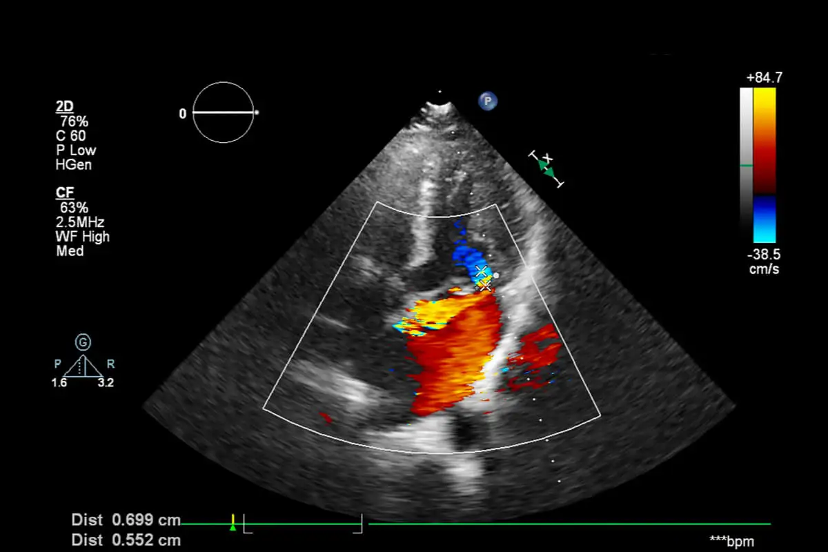
As we mentioned before, fetal echocardiography is a non-invasive method of imaging the baby’s heart. The procedure gives a two- and three-dimensional image of the 4 chambers of the heart and outflow ducts, as well as through the spectral and color Doppler effect allows to evaluate blood flows.
On the other hand, it allows experts to evaluate the heart rate and hemodynamic condition of the baby. That is, the “moving image” of the functioning of the heart muscle.
In simpler terms, we would say that there are two forms of images that the study will provide:
- Two-dimensional or 2D echocardiogram: Although it receives this name, it’s a combination of 2D and 3D. The physician observes the anatomy and functioning of the heart’s chambers as blood enters and exits.
- EchoDoppler: Through the use of a physics effect, the device measures the speed with which the blood passes from one chamber to another and into the vessels. It thus makes it possible to evaluate the dynamics of the fluid between the different parts of the heart and the real-time functioning of the valves. According to experts, it’s one of the analyses that is almost always requested in the context of a high-risk pregnancy.
When should it be performed?
Fetal echocardiography can be performed during the first trimester of gestation, between weeks 11 and 14. Therefore, the images wouldn’t provide useful information.
In turn, the transabdominal route for ultrasound is recommended between 18 to 24 weeks of gestation (second trimester). After 30 weeks, the bony structures of the fetus, as well as the air in the lungs, prevent a good view of the heart.
In summary, the ideal times for the study are the 11-14 and 18-24 week intervals.
Learn more: Everything You Need to Know About Fetus in Fetu
When is fetal echocardiography performed?
As mentioned, fetal echocardiography isn’t performed in all pregnancies. It’s indicative in pregnancies with high suspicions of cardiac malformations. Therefore, it’s assumed that this study would certify an initial suspicion of something that was visualized in the usual prenatal ultrasound.
It should also be performed in women with certain risk factors, such as the following:
- Maternal phenylketonuria
- Known genetic alterations
- Family medical history of congenital cardiac malformations
- Maternal metabolic diseases (gestational diabetes and arterial hypertension) or inflammatory diseases (lupus)
- Maternal viral infections: rubella, toxoplasmosis, cytomegalovirus, measles, and human immunodeficiency virus (HIV)
- Maternal consumption of cardiac teratogens: indomethacin, steroids, tobacco, alcohol, amphetamines, progestins, and anticonvulsants
- Highly complex reproductive techniques by egg donation or intracytoplasmic sperm injection
- Twin pregnancies, especially those that share a single placenta (monochorionic)
- History of pregnancy with congenital malformations
If an increased nuchal translucency appears in a routine ultrasound scan, then a fetal echocardiogram should be performed. This is stated in a 2016 review. It should be performed whenever a sonographer finds something that raises concerns, not only in the shape of the heart, but also regarding the rhythm of the heartbeat.
Learn more about: What is Congenital Hypothyroidism?
What’s the procedure like?
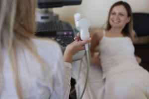
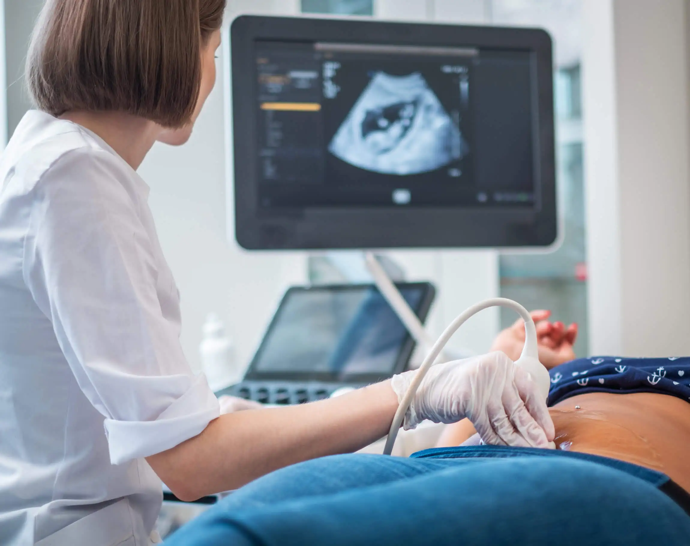
The procedure for fetal echocardiography is practically the same as for a prenatal ultrasound. It’s performed in a darkened room, with the pregnant woman lying on a bed. Most often, the transabdominal technique is used, but, in the first trimester, a transvaginal technique may be performed.
In the case of the transabdominal procedure, a conductive gel is placed on the abdomen that allows the sound wave to travel from the device to the baby’s heart and back. This allows the images to be obtained on a monitor.
The procedure usually takes no more than 30 minutes to an hour, but will depend on the position of the baby, the structures to be explored, and the possible malformations found, as well as the gestational age and experience of the performing physician.
The results will be given the same day if there’s an urgent need. However, sometimes more time will be needed for a detailed evaluation by specialists.
When fetal echocardiography detects a cardiac malformation, it will be the pediatric cardiologist who will explain in detail the findings and the steps to follow to the parents.
It isn’t painful and poses no risk
Fetal echocardiography is a harmless, non-invasive, and painless procedure. The mother may feel a little pressure when placing the transducer, but nothing more. It also requires no prior preparation.
The importance of screening with fetal echocardiography
Fetal echocardiography detects up to 90% of congenital heart disease. Although it is intended for early detection, sometimes some defects cannot be certified until delivery. These include mild valvular problems or small communications between heart chambers.
Congenital heart defects are the most frequent congenital malformations and are also a frequent cause of death in the neonatal stages and in the first year of a baby’s life. For this reason, fetal echocardiography has now been proposed as part of a screening test for all pregnant mothers.
Even so, it isn’t yet part of the tests that are required every trimester in a normal pregnancy. If your obstetrician or sonographer has any concerns, then they will request this procedure.
All cited sources were thoroughly reviewed by our team to ensure their quality, reliability, currency, and validity. The bibliography of this article was considered reliable and of academic or scientific accuracy.
- De Rubens J. (2005). La ecocardiografía fetal como método de diagnóstico temprano. Acta Pediátrica de México, 26, 5. repositorio.pediatria.gob.mx:8180/bitstream/20.500.12103/1585/1/ActPed2005-38.pdf
- Finch A. (2005). La importancia de la ecocardiografía fetal en la detección y el manejo de las malformaciones cardíacas congénitas. Problemas relevantes en cardiología. Rev Esp Cardiol, 59, 2. https://www.sciencedirect.com/science/article/abs/pii/S0300893206752368
- Góngora O. (2020). Importancia de la ecocardiografía fetal en el diagnóstico de malformaciones congénitas. CorSalud, 12, 4. https://www.medigraphic.com/pdfs/corsalud/cor-2020/cor204t.pdf
- Grilloni, M. M., Uyuni, N., García Pérez, D. A., Papa, F. A., Bobadilla, S. A., & Mora, A. J. (2018). Utilidad y aplicación del ecodoppler en el embarazo de alto riesgo. Hosp. Aeronáut. Cent, 95-104. https://pesquisa.bvsalud.org/gim/resource/fr/biblio-1021165
- Hospital Clinic, Hospital Sant Joan de Déu, Universitat de Barcelona. (S.F.). Ecocardiografía fetal. Protocolo: ecocardiografía fetal. https://repositorio.upn.edu.pe/bitstream/handle/11537/32314/ecocardiografia%20fetal.pdf?sequence=1
- Muñoz, H., Enríquez, G., Ortega, X., Pinto, M., Hosiasson, S., Germain, A., … & Cortés, F. (2023). Diagnóstico de cardiopatías congénitas: ecografía de cribado, ecocardiografía fetal y medicina de precisión. Revista Médica Clínica Las Condes, 34(1), 44-56. https://www.sciencedirect.com/science/article/pii/S0716864023000019
- RuotiCosp, M. (2016). Manejo del feto con translucencia nucal aumentada. Anales de la Facultad de Ciencias Médicas (Asunción), 49(2), 49-67. http://scielo.iics.una.py/scielo.php?pid=S1816-89492016000200006&script=sci_arttext
This text is provided for informational purposes only and does not replace consultation with a professional. If in doubt, consult your specialist.


