Hemispherectomy and the Postoperative Period
A hemispherectomy is a surgical procedure used to treat various seizure disorders. Typically, doctors perform it when these disorders don’t respond adequately to other less radical treatments.
The first hemispherectomy was performed on a dog in 1888. The first reference to this procedure performed in humans dates back to 1923. In the 60s and 70s, several interventions yielded poor results.
These days, things have changed. Doctors often opt for an anatomical hemispherectomy instead of a functional hemispherectomy. This is a more accurate and less invasive intervention. The success rate is much higher than in the past.
In this article, we’ll take a closer look at the modern forms of this procedure.
What’s a Hemispherectomy?
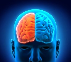
A hemispherectomy is a neurosurgical procedure that involves the removal of one of the cerebral hemispheres.
Sometimes, the surgeon removes the left hemisphere. Other times, they remove the right hemisphere. Doctors mainly perform this procedure in extreme cases and on children aged 5 to 10.
This type of intervention is primarily an anti-convulsive treatment. However, it’s also useful for patients with neurological deficits and in severe cases of serious head trauma.
The entire cerebral hemisphere is removed in most interventions. However, sometimes only part of it is removed. This is called a functional hemispherectomy. In such cases, if doctors leave a small portion of damaged tissue behind, the seizure disorder may manifest again.
Indications
Generally speaking, medical professionals may indicate a hemispherectomy for patients who suffer from continuous and daily seizures that haven’t responded to other medical treatments or other less invasive surgical procedures.
Medical professionals recommend it in the following cases:
- Children with hemiplegia. This is only for children over the age of four that suffer from seizure and/or mental disorders, and after checking for two years that the patient hasn’t responded to drug treatment.
- Sturge-Weber syndrome. This is a neurocutaneous disorder characterized by a facial birthmark in the area of the trigeminal nerve. Doctors may recommend this type of surgery when crises start at an early age and the disorder affects the entire hemisphere.
- Rasmussen’s encephalitis. This brain disorder causes chronic, progressive encephalitis. It is best to receive early intervention.
- Hemimegalencephaly (HME). This is a rare inflammatory neurological disease that causes severe seizures. Medical professionals still don’t agree on if surgery is the best option for patients who suffer from this disorder.
- Cortical development malformations.
Read: Revolutionary Therapy Gives a Man in a Coma his Consciousness Back
Characteristics of the Procedure
Overall, there are four types of hemispherectomy.
The four types are:
- Anatomical hemispherectomy
- Hemidecortication
- Functional hemispherectomy
- Modified functional hemispherectomy
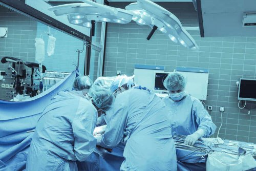
Typically, doctors use anesthesia for the procedure.
The surgeon starts by shaving the patient’s head and marking the incision lines. Then, the medical professional cuts to expose the dura mater. Then, they remove to reach the brain.
After, they carefully mark the area they must remove. They then do so and cauterize the blood vessels. They place a drainage device. Finally, they place the dura mater and scalp and close the incision with staples.
Hemispherectomy Postoperative Period
Unfortunately, the postoperative period for this procedure is painful.
Typically, doctors leave the drainage tube for 3 to 4 days. After, the medical professional evaluates the patient and decides if they should remove it or not. Before removing it, they must perform diagnostic tests to determine whether there’s bleeding or hemorrhage.
The main complications are hemodynamic instability, hypothermia, and hypo- or hyperkalemia. Normally, the doctors control and prevent them very well.
Seizures during the postoperative period are another serious complication. Typically, about half of the patients develop hydrocephalus. Meanwhile, almost all patients develop aseptic meningitis.
Also, there’s evidence that some complications may manifest sometime later.
Read on: What is Meningitis?
Nevertheless, the mortality rate is quite low and ranges between 4% and 6%.
Between 70% and 85% of patients who undergo a hemispherectomy stop suffering from seizures. Meanwhile, about 10-20% significantly improve their quality of life.
All cited sources were thoroughly reviewed by our team to ensure their quality, reliability, currency, and validity. The bibliography of this article was considered reliable and of academic or scientific accuracy.
- Obrador Alcalde, S. (1951). Hemisferectomía en el tratamiento de las convulsiones de la hemiplejía infantil por hemiatrofía cerebral. Arquivos de Neuro-Psiquiatria, 9(3), 191-197.
- Alcalá-Cerra, G., Paternina-Caicedo, Á., Díaz-Becerra, C., & Gutiérrez-Paternina, J. J. (2013). Control de las crisis epilépticas con la hemisferectomía cerebral en adultos: revisión sistemática y metaanálisis con datos de pacientes individuales. Neurocirugía.
- Meneses, M. S. de, Kondageski, C., Santos, H. N. L. dos, Kowacs, P. A., Coelho, G. C., Gadens, G., … Simão, C. (2012). The usefulness of neuronavigation in functional hemispherectomy. Journal of Epilepsy and Clinical Neurophysiology.
This text is provided for informational purposes only and does not replace consultation with a professional. If in doubt, consult your specialist.


