Electroencephalogram: The Process, Results, and Common Questions

Reviewed and approved by the nurse Leidy Mora Molina
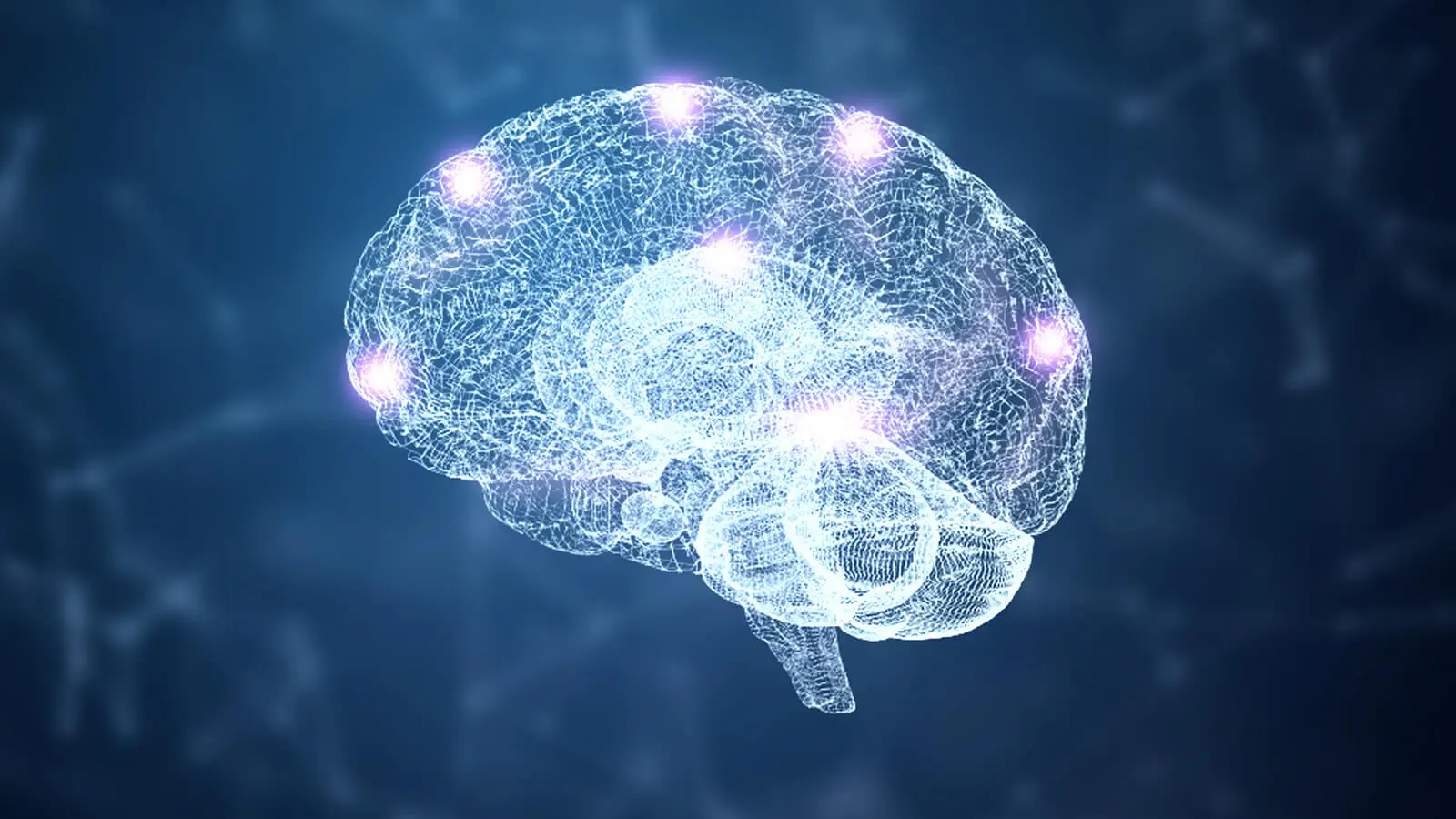
The brain is made up of a large and complex network of specialized cells called neurons. It communicates through electrical stimuli that can be analyzed by means of the electroencephalogram.
The health of the human brain and the entire central nervous system (CNS) can be assessed through a large number of tests. In this way, it’s possible to detect structural abnormalities and disorders of nerve function in time. Electroencephalography, tomography, and magnetic resonance imaging are some of the most common tests.
Electroencephalogram is the method of choice for the diagnosis of seizures and epilepsy, according to specialists. This medical tool has saved millions of lives. However, its usefulness goes far beyond diagnosis.
What is the electroencephalogram?
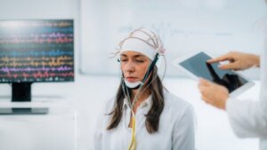
This is a functional exploration technique that measures the brain’s electrical activity in real time. According to studies, it was Hans Berger who coined the term electroencephalogram (EEG) in 1929 to describe the recording of brain electrical fluctuations captured by electrodes attached to the scalp.
Neurons are always active, transmitting electrical impulses throughout the CNS. EEG captures and amplifies these electrical signals, representing them in wavy lines that define the activity of the different brain regions.
In most cases, an EEG is performed in the basal state and subject to activation methods, such as hyperventilation or visual stimulation. In fact, some professionals recommend obtaining a detailed brain recording during sleep. In addition, 24-hour follow-up EEGs are available.
This test results in normal and abnormal patterns that allow the diagnosis of injuries or characteristic disorders, such as seizures. For this reason, it’s a complementary study widely used in neurological consultation.
Why is this test performed?
It’s of medical interest to evaluate the state of brain electrical activity in those persons with suspected episodic or persistent nervous disorders. Among the most frequent indications for electroencephalograms are the following:
- Alteration of higher functions, such as memory and consciousness
- Epilepsy or other convulsive syndromes
- Sleep disorders, such as insomnia
- Encephalitis and other CNS infections
- Cerebrovascular disease (CVD)
- Monitoring during brain surgery
- Traumatic brain injury
- Cranial tumors
- Alzheimer’s disease
Likewise, an EEG is useful to confirm brain death in patients in a deep coma. Likewise, it offers data of interest in drug-induced anesthesia.
We think you may also enjoy reading this article: What is a Brain Aneurysm? Learn About Emilia Clarke’s Condition During Games of Thrones
Possible risks and contraindications
In general, an EEG is a fairly safe technique and doesn’t cause any type of pain or adverse reaction. Controlled methods are sometimes used to induce seizures, such as light stimulation or hyperventilation. However, the specialist is trained to provide medical attention if necessary.
Preparations for an EEG
There are several recommendations that should be followed before performing an EEG. In this way, we ensure that the test is performed correctly and without errors in the results.
In this regard, preparations for the EEG include the following:
- Wash your hair the night before or several hours prior to the study.
- Avoid using hair conditioners, gels, oils, creams, or hairsprays, as they may hinder the adhesion of the electrodes.
- In the case of hair extensions, ask your healthcare provider for instructions.
- Don’t change or discontinue any regular medication without your doctor’s indication. Consult your healthcare provider for further information.
- Avoid caffeinated foods or beverages 6 to 8 hours before the test.
- Sleep less than usual in the case of an EEG during sleep.
- Don’t take energizers or other products to stay awake, especially if you need to sleep during the test.
How is the EEG performed?
This test is performed by a professional EEG technician in a medical center, private office, or laboratory. He/she will be in charge of guiding the patient through the whole process in a safe and simple manner.
During the test
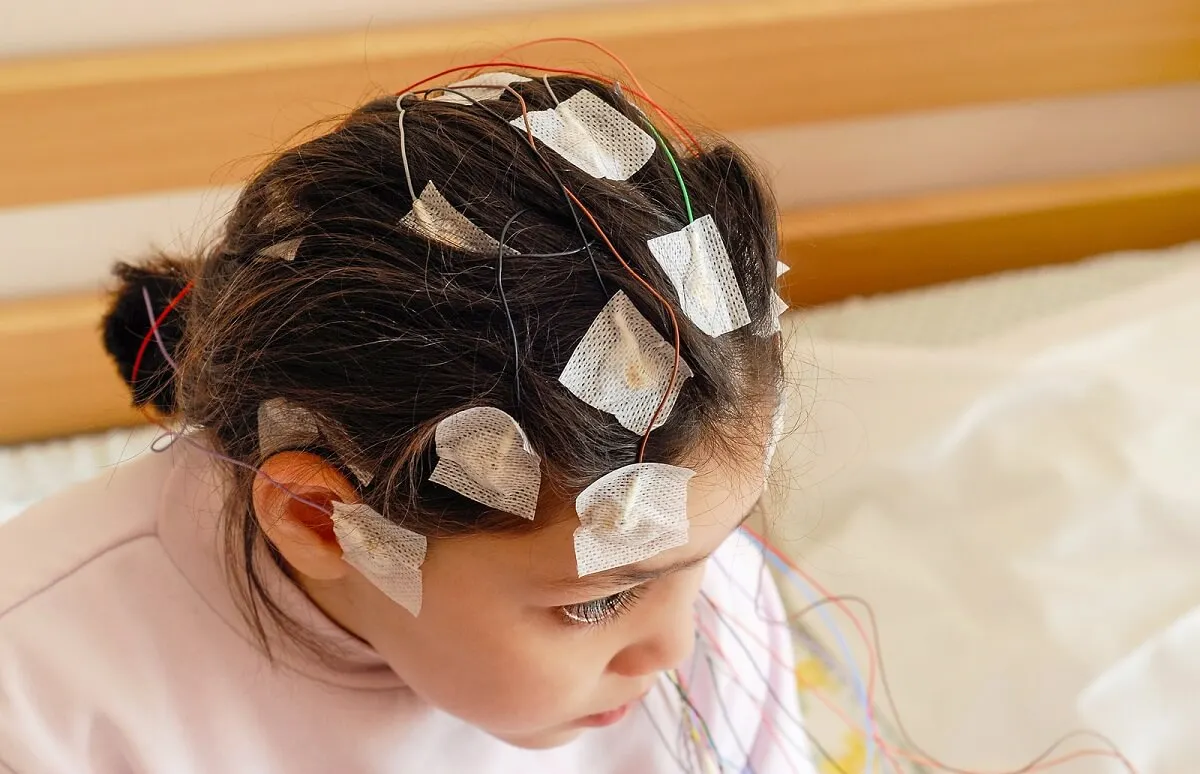
The person to be evaluated must lie down on a stretcher or reclining chair. Then, a technician will be in charge of measuring the different diameters of the skull and marking the points where the electrodes will be placed. These discs don’t produce any type of pain and are in charge of recording brain activity.
In general, the electrodes are placed on the scalp using a special adhesive. Sometimes, caps that include the electrodes are used. The electrodes will be connected by wires to an instrument that will capture and amplify the electrical signal.
During the test, the person being tested must remain relaxed and with his or her eyes closed. Occasionally, the technician may ask the person to open and close the eyelids, take a quick, deep breath, perform a calculation, or look at a bright light. In addition, the patient may also be asked to sleep during the evaluation.
In most cases, the body movements during the EEG are captured on video. In this way, the physician can combine the brain wave recording with these images to obtain a more accurate diagnosis.
On the other hand, an ambulatory EEG may be indicated for those who require more prolonged monitoring. This unit will accompany the patient throughout the day and will keep the brain’s electrical recordings while the usual activities are performed.
Like this article? You may also like to read: The Modular Theory of Mind: How Does the Human Brain Work?
After the test
The basic EEG usually lasts 20 to 40 minutes, once the electrodes are in position. However, as mentioned earlier, some tests may require the patient to sleep, which lengthens the test.
At the end of the EEG, the technician will remove the electrodes from the scalp. Some people may require sedatives to induce sleep, so a friend or family member may need to accompany them home after the test. If you don’t consume any type of sedative, you can resume your daily activities as normal.
Results of the electroencephalogram
This test provides a printed record of brain activity, represented by waves that are drawn with different frequencies and amplitudes. EEG characteristics vary according to the state of consciousness. In this sense, the waves are usually faster when the patient is awake and slower during sleep phases.
Health professionals are the only ones qualified to interpret an EEG and provide an accurate diagnosis. In general, the presence of broad, sharp waves may indicate a seizure syndrome, such as epilepsy. Bleeding, tumors, and brain aneurysms are other common causes of abnormal findings.
If you have any concerns, don’t hesitate to consult a physician specializing in neurology.
EEG: A useful tool for assessing brain health
The EEG is a safe and useful test for evaluating the health of the brain and the entire central nervous system. Early diagnosis of neurological diseases improves the quality of life of many people and prevents long-term complications. Its administration and interpretation must be done by health professionals.
All cited sources were thoroughly reviewed by our team to ensure their quality, reliability, currency, and validity. The bibliography of this article was considered reliable and of academic or scientific accuracy.
- Viloria Alebesque A, López Bravo A, Bellosta Diago E, Santos Lasaosa S, et al. Utilidad del electroencefalograma en el manejo de la epilepsia en el Servicio de Urgencias. Neurología. 2020; 35(4):238-244.
- Ramos F, Morales G, Egozcue S, Pabón R, et al. Técnicas básicas de electroencefalografía: principios y aplicaciones clínicas. An. Sist. Sanit. Navar. 2009; 32 (Supl. 3): 69-82.
- Monge-Pereira E, Molina-Rueda F, Rivas-Montero FM, Ibáñez J, Serrano JI, et al. Electroencephalography as a post-stroke assessment method: An updated review. Neurologia. 2017 Jan-Feb;32(1):40-49.
- Iriarte J, Urrestarazu E, Alegre M, Martín B, et al. Vídeo-electroencefalografía: una necesidad. An Sist Sanit Navar. 2009;32 (Suppl 3):83-92.
- Berenguer-Sanchez MJ, Gutierrez-Manjarrez F, Senties-Madrid H, Estanol-Vidal B. Variantes normales o de significado incierto en el electroencefalograma. Rev Neurol. 2012 Apr 1;54(7):435-44.
This text is provided for informational purposes only and does not replace consultation with a professional. If in doubt, consult your specialist.


