Basal Cell Carcinoma: The Most Common Skin Cancer

Reviewed and approved by the nurse Leidy Mora Molina
Skin cancer is one of the most common forms of cancer worldwide. In particular, basal cell carcinoma and squamous cell carcinoma are the most frequent types of skin cancer.
In general, all neoplastic diseases are the result of the abnormal and uncontrolled growth of a group of cells. This type of cancer, also called “basal cell carcinoma,” begins in the basal cells of the deepest layer of the epidermis, which are responsible for skin regeneration.
The American Cancer Society estimates that 8 out of 10 cases of skin cancer are basal cell carcinomas, with an incidence in the United States of more than 5 million people affected. Early diagnosis and a timely approach determine a better prognosis and quality of life.
Common symptoms of basal cell carcinoma
Basal cell carcinoma is a slow-growing and painless type of neoplasm, which in some cases, goes easily unnoticed. Likewise, it tends to develop more frequently in sun-exposed areas of the body.
Studies affirm that 90% of cases occur in the head and neck. Of these, 10% occur in the eyelids, as this is the most frequent type of malignant eyelid tumor. On the other hand, a small number of cases occur in areas of skin protected from the sun, such as the genitals.
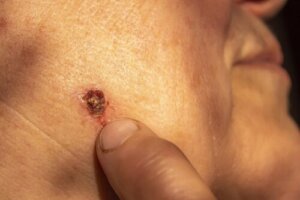
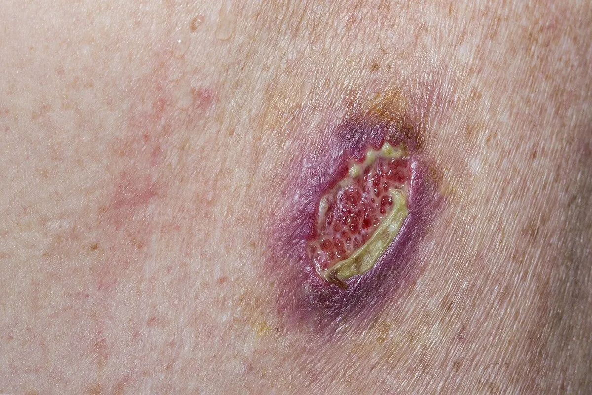
However, most of the time, it manifests as a lump, a bump, or a sore on the skin that grows slowly and doesn’t heal. However, the characteristics of the lesion vary depending on the type of basal cell cancer. Some of the most common lesions are as follows:
- A shiny, pearly, translucent lump with a tendency to bleed and crust
- A brown, bluish, or blackish-brown bump with translucent edges
- Scaly, red plaque with raised edges
- A whitish, waxy patch with poorly defined edges, similar to a scar
Small dilated vessels or telangiectasias may appear over time, accompanying the skin lesions. The latter increases the risk of bleeding and ulcer formation.
Other less common types present as small, superficial pink or red patches. In addition, a thick, fleshy, pearly-colored lesion on the skin may also indicate its presence.
We think you may also enjoy reading this article: Skin Cancer: Warning Signs, Causes And Symptoms
The causes of basal cell carcinoma
Basal cell cancer originates at the level of the basal cell layer of the epidermis, the latter being the most superficial layer of the body. Basal cells are responsible for producing new skin cells and replacing old cells that eventually slough off the skin.
Mutations in the genetic code or DNA of these cells lead to their abnormal multiplication. Eventually, uncontrolled growth of the basal cell layer promotes the formation of a visible skin tumor.
Research has shown that ultraviolet (UV) radiation is the main trigger for mutations in the DNA of skin cells. In general, the sun’s rays are the major source of UV radiation, as are tanning beds. However, there are some forms of basal cell carcinoma that are not associated with this cause.
Risk factors
It often occurs in older adults as a result of its slow and inconspicuous growth. Studies estimate that this cancer is 100 times more frequent between the ages of 55 and 75 than in those under 20 years of age.
On the other hand, the incidence is usually higher in males. In fact, it occurs 30% more in men than in women. This is related to a greater source of occupational exposure to the sun. Other risk factors associated with its appearance are the following:
- Light skin
- Light eyes and blond or reddish hair
- Repeated exposure to x-rays or other sources of radiation
- Having many moles and freckles
- A family history of skin cancer
- A personal history of skin tumor lesions
- Sunburns at an early age
- Outdoor occupations
- Depressed immune system
- The use of immunosuppressive drugs
- Exposure to arsenic
In turn, there are some hereditary syndromes that increase the risk of skin cancer. Such is the case of nevoid basal cell carcinoma syndrome – also called Gorlin-Goltz disease – and xeroderma pigmentosum.
Like this article? You may also like to read: 6 Skin Cancer Symptoms You Should Not Ignore
Associated complications
In general, the greatest fear of any neoplasm is its dissemination to other organs, or metastasis. When a basal cell carcinoma grows on the face – near the eyes, ears, or mouth – the consequences of spread can be fatal. Fortunately, it rarely metastasizes.
As for the likelihood of recurrence, in many cases, there’s a high risk, even after a successful treatment plan. In addition, basal cell carcinoma increases the risk of other skin malignancies, such as squamous cell cancer.
Diagnosis
Diagnosis of skin cancer requires a comprehensive evaluation by a medical specialist. During the examination, questions may be asked about possible skin changes, family history of the disease, and personal history of other conditions.
It’s crucial to report any unusual symptoms or signs to the treating physician. Physical examination is vital in identifying basal cell carcinoma, as most are able to recognize it with the naked eye. In addition, it’s important to evaluate other regions of the body for significant changes in the skin.
If there’s suspicion of a tumor lesion, the specialist may ask to take a sample of the affected tissue. A biopsy is the method of choice for the definitive diagnosis of skin cancer, according to studies. Its choice depends on the size and type of lesion.
Treatment of basal cell carcinoma
Treatment of basal cell cancer is focused on removing the lesion from the skin and preventing its spread to other tissues. Surgical excision is usually the most common method used to remove it. This involves cutting the lesion and a margin of healthy skin for later analysis.
For some types of larger tumors or recurrent lesions, Mohs micrographic surgery may be necessary. This is a microscopically guided procedure in which the lesion is removed and examined layer by layer until no tumor cells remain.
On the other hand, there are several techniques for cases in which surgery is not possible or not recommended. Curettage and electrodesiccation allow the lesion to be removed by scraping and then sealed with heat. Other procedures include radiotherapy, photodynamic therapy, and cryosurgery.

Recommendations to prevent basal cell cancer
Currently, there are several habits to reduce the risk of developing basal cell carcinoma. Some recommendations to prevent this type of skin cancer are the following:
- Avoid sun exposure during the strongest hours, between 10:00 am and 4:00 pm.
- Use sunscreen of at least 30 SPF every day.
- Reduce visits to tanning salons.
- Wear protective clothing when outdoors, such as hats, long pants, and long-sleeved shirts.
- Create a skin check-up routine.
- Consult a physician if you notice any lumps or strange lesions.
Early detection improves the prognosis of basal cell carcinoma
As you can see, basal cell carcinoma is the most common form of skin cancer presentation. Fortunately, most of these lesions are cured when treated early. Rarely do they spread beyond the affected site.
So, be sure to seek professional attention for any skin lesions with changes in size, color, or appearance.
All cited sources were thoroughly reviewed by our team to ensure their quality, reliability, currency, and validity. The bibliography of this article was considered reliable and of academic or scientific accuracy.
- Álvarez Castillo A, Rodríguez Alfaro J, Salas Boza A. Revisión sistemática del carcinoma basocelular. Revista Medica Sinergia. 2020;5(5):e483.
- Samela P, Tosi V, Cervini A, Bocian M, et al. Síndrome del nevo basocelular: experiencia en un hospital pediátrico. Actas Dermo-Sifiliográficas. 2013;104(5):426-433.
- Castañeda Gameros P, Eljure Téllez J. El cáncer de piel, un problema actual. Rev. Fac. Med. (Méx.). 2016 ; 59( 2 ): 6-14.
- Urrego-Rivera F, Faura-Berruga C. Diagnóstico diferencial del carcinoma basocelular pigmentado. Rev Clin Med Fam. 2015; 8( 2 ): 166-170.
This text is provided for informational purposes only and does not replace consultation with a professional. If in doubt, consult your specialist.


