The Types of Synovial Joints
Synovial joints provide a connection between two bones. They’re made up of a cartilage-lined cavity filled with fluid. In this article, we’ll take a closer look at what these joints are, where they’re located, and their function.
The Characteristics of Synovial Joints
First of all, the joints have a layer of cartilage that covers the articular surfaces of skeletal elements. Thus, the bone surfaces don’t contact each other directly. When these joints are visible on X-rays, there seems to be ample space between adjacent bones.
Plus, the second characteristic of synovial joints is the presence of an articular capsule. Basically, it consists of an inner synovial membrane and an outer fibrous membrane.
The synovial membrane
This membrane lines the inner surface of capsules of synovial joints and tendon sheaths. Overall, it makes contact with the fibrous membrane and the synovial fluid lubricant.
The synovial membrane is highly vascularized and produces synovial fluid. Then, this builds up in the joint cavity and lubricates the joint surfaces.
Also, closed synovial membrane sacs appear outside the joints. This is where they form bursae or tendon sheaths. The sacs often form between structures such as tendons and joints, tendons and bone, or skin and bone and reduce friction when one structure moves over another. Also, tendon sheaths surround tendons and reduce friction.
Discover: Anti-inflammatory Cayenne Pepper Ointments for Joint Pain
The fibrous membrane
Next, the fibrous membrane is made up of dense irregular connective tissue. Overall, it surrounds and stabilizes the joint.
Parts of the fibrous membrane may thicken to form ligaments that further stabilize the joint. The ligaments that are located outside the capsule often provide additional reinforcement.
Other characteristic structures of synovial joints
Another characteristic of synovial joints is the presence of additional structures within the area encompassed by the synovial membrane. These include:
- Articular disks. Usually, these are made up of fibrocartilage. They absorb compressive forces, adjust to joint surface changes during movement, and increase range of movements that can occur in the joints.
- Articular fat pads. These are often located between the synovial membrane and capsule. Also, they enter and leave these areas as the articular contour changes during movement.
Read on to learn more: Joints: Their Importance and Necessary Supplements
Types of synovial joints
The body has the following types of synovial joints:
- Plane joints. Overall, these allow sliding movements when a bone moves over another. One example is the acromioclavicular joint, which is located at the top of the shoulder.
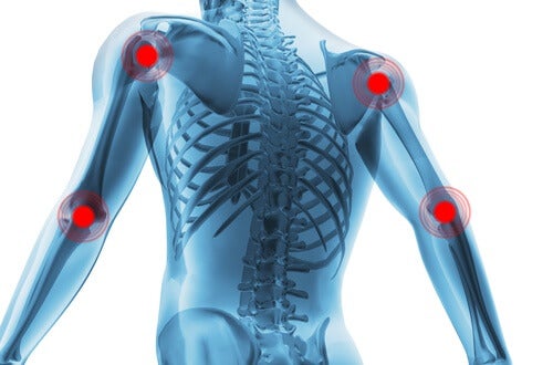
- Hinge joints are joints that allow flexion and extension in one plane. One example is the elbow.
- Pivot joints allow movement around a shaft, meaning they regulate rotation movements. One example is the distal radioulnar joint.
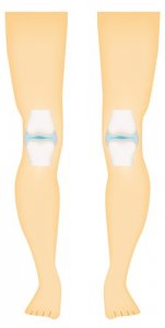
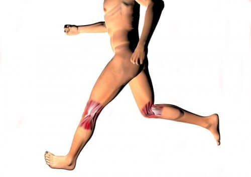
- Compound or bicondyloid joints. These joints allow movement between an axis, with limited rotation in a second axis. They consist of two convex condyles. An example of this type of joint is the knee joint.
- Condyloid joints allow movement within two perpendicular axes and passive or secondary movement on a third axis. Thus, they allow flexion, extension, abduction, and adduction movements (circumduction). This is the case of the wrist joint.
- Saddle joints. These allow the same movements as the condyloid joints but allow greater movement. This name derives from their saddle shape. They provide flexion, extension, abduction, and adduction movements (circumduction).
- Ball joints allow all movements except gliding. They provide flexion, extension, abduction, rotation, and adduction movements (circumduction). The hip is an example of this type of joint.
Conclusion
If we didn’t have joints, the bones of our body couldn’t move. Thanks to our joints, we can make different movements such as bending, rotating, and flexing our arms and legs!
All cited sources were thoroughly reviewed by our team to ensure their quality, reliability, currency, and validity. The bibliography of this article was considered reliable and of academic or scientific accuracy.
-
Padrós Soler, G., Galán Ortega, A., Guillén Campuzano, E., Hortas Nieto, M. L., Marín Soria, J. L., Muñoz Pérez, M., & Noguera Bennaser, A. (2004). Recomendaciones para el estudio del líquido sinovial. Química Clínica.
-
Iturriaga, V., Mena, P., Oliveros, R., Cerda, C., Torres, D., & del-Sol, M. (2018). Importancia del Líquido Sinovial en la Articulación Temporomandibular y sus Implicancias en la Patología Articular. International Journal of Morphology. https://doi.org/10.4067/s0717-95022018000100297
-
Aguado Jódar, X. (1992). Diferencias y similitudes entre el rozamiento deslizantes y de giro. Fuente: Serway R. A.. Física. Editorial McGraw-Hill.
This text is provided for informational purposes only and does not replace consultation with a professional. If in doubt, consult your specialist.


