Pericardial Effusion: Diagnosis and Treatment
Pericardial effusion is a condition that involves fluid accumulation around the heart. Sometimes, it’s related to an illness. However, other times there needs to be specific monitoring and evaluation to establish its cause. In some cases, it may not be possible to identify a cause. In fact, it can even become chronic without causes serious problems. However, it’s very important to get treatment.
Typically, treatment focuses on solving the problem that causes the pericardial effusion and managing its symptoms. However, if the cause is unknown, the clinical management is the same as pericarditis.
What’s pericardial effusion?

This condition is the accumulation of fluid between the membranes of the heart.
Pericardial effusion is an abnormal accumulation of fluid in the pericardial cavity. The pericardium is made of two layers: a serous layer and a fibrous layer. The space between these two layers is the pericardial cavity. Normally, it contains up to 50 ml of serous fluid. When there’s an inflammatory or infectious process, liquid production increases and pericardial effusion occurs.
Also, this can manifest because of a decreased reabsorption of the liquid. Generally, this occurs due to increased systemic venous pressure. In turn, the increase in pressure typically occurs due to congestive heart failure or pulmonary hypertension.
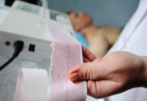
Diagnosis
The clinical presentation of pericardial effusion depends on the speed with which fluid accumulates. Typical symptoms are shortness of breath and chest pain. Nausea, dysphagia, hoarseness, and hiccups are also common symptoms.
When a medical professional suspects pericardial effusion, they may request one or more of the following tests:
- Echocardiogram. It allows the doctor to detect the magnitude of the effusion and evaluate heart function.
- Electrocardiogram. This test can detect potential blockage patterns.
- Chest X-rays. These allow medical professionals to establish the magnitude of pericardial effusion.
The most widely used diagnostic test is echocardiogram. However, computed tomography (CT) and magnetic resonance (MR) offer a wider field of view. Nevertheless, due to availability and costs, medical professionals don’t resort to them as frequently.
In any case, echocardiographic evaluation allows the determination of five key variables: size, duration, distribution, composition, and hemodynamic effects. They used these to establish the cause of the pericardial effusion to decide the appropriate course of treatment.
Discover more in this article: Cream of Garlic Soup for Heart Health
Pericardial effusion treatment

Typically, medical treatment indicated for these cases is nonsteroidal anti-inflammatory drugs.
Overall, the pericardial effusion treatment directly depends on several factors. These include the amount of accumulated liquid, the existence of cardiac tamponade, and the cause. In general, the problem goes away as soon as doctors can treat the cause.
The first step in managing pericardial effusion is to assess its size. Also, doctors must define its hemodynamic significance and establish possible associated diseases. There’s an underlying disease in approximately 60% of cases. If there’s no tamponade or considerable risk of it occurring, most doctors indicate bed rest and anti-inflammatory treatment. Often, they also prescribe colchicine and corticosteroids.
However, if there’s a risk of tamponade or high risk that the effusion will progress, doctors should perform a pericardiocentesis. When it’s not possible to perform this procedure or it fails, open surgical drainage is the next step. This should include a biopsy and the creation of a pericardial window.
This article may interest you: First Aid: How to Respond During a Cardiac Emergency
Monitoring and prognosis
In general, idiopathic pericardial effusion and pericarditis have a good prognosis. Thus, the risk of complications is very low. Cases of chronic idiopathic pericardial effusion have a probability of between 30% and 35% of scaling to cardiac tamponade. In the other type of effusion, the prognosis depends primarily on the cause that produces it and its size. In approximately one-third of the total cases, those greater than 10 mm worsen and evolve to tamponade.
However, it’s advisable to monitor moderate Idiopathic effusions with an echocardiogram every six months. If severe, the patient should be monitored every three months. In the case of non-idiopathic effusions, monitoring will depend on the disease that caused the pericardial effusion.
All cited sources were thoroughly reviewed by our team to ensure their quality, reliability, currency, and validity. The bibliography of this article was considered reliable and of academic or scientific accuracy.
- CLAVERÍA, C., VERGARA, L., NEGRÓN, S., & ZELADA, P. (2009). Derrame pericárdico, enfrentamiento clínico. Revista chilena de pediatría, 80(3), 267-273.
This text is provided for informational purposes only and does not replace consultation with a professional. If in doubt, consult your specialist.


