A Hypoechoic Nodule: What Is It and How to Identify One?
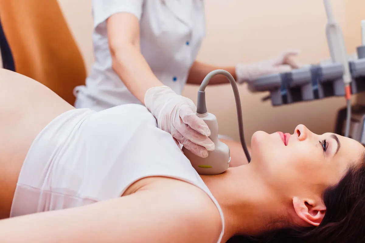
Ultrasonography, or ultrasound, is a widely used imaging study to explore the interior of the human body. This technique makes it possible to identify hypoechoic, hyperechoic, isoechoic, and anechoic structures. Are you interested in knowing how to identify a hypoechoic nodule? We’ll tell you here.
In general, nodules are rounded lesions, similar to a lump, produced by the accumulation of liquids and tissues. They can occur on the outside or inside of the body. The breasts, liver, thyroid, uterus, and lymph nodes are some of the most common areas for nodule formation.
In this sense, ultrasound allows early localization of any hypoechoic nodules that may be present in any organ. From the characteristics of the image, it’s possible to identify whether the lesion is benign or whether it could have a malignant component.
Health professionals are the only ones qualified to evaluate these lesions.
What is a hypoechoic nodule?
The term echogenicity describes the capacity of an organ or other structure to reflect or bounce back ultrasound signals. The less dense the tissue, the lower its capacity for reflection; consequently, it will have a lower echogenicity.
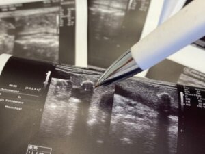
A hypoechoic nodule, also called hypoechogenic, is a mass formed by tissues, fat, or liquids of low density. For this reason, the ultrasound signal penetrates more easily, offering a dark gray image that differs from the surrounding structures.
Studies define a hypoechoic structure as one that generates few echoes and has low density. Hypoechoicity makes it possible to differentiate abnormal lesions that are immersed in any tissue.
You may be interested in reading: Thyroid Biopsies: Everything You Need to Know
Types of nodules
Nodules can originate anywhere in the body. It’s possible to classify these lesions taking into account their composition and content:
- Cystic: These are nodules of liquid content covered by a capsule. Most hypoechoic nodules are of this type.
- Solid: A thick dense cellular mass.
- Mixed: These are nodules that include solid structures and some liquid areas.
What other types of images does ultrasound offer?
This imaging test allows the detection of different forms of echogenicity, apart from hypoechoic structures. In this sense, we can also find the following:
- Hyperechoic or hyperechogenic: Thia is a highly reflective white-based image, typical of calcifications and bones.
- Isoechoic or isoechogenic: A light gray homogeneous structure, common in tendons.
- Anechoic or anechogenic: A black-based image without reflective capacity. It’s typical of liquid-containing nodules.
When is a hypoechoic nodule malignant?
Nodules can be benign or malignant, depending on their ability to penetrate the surrounding tissue and spread to other organs. In most cases, cystic lesions have a lower risk of malignancy. However, this isn’t true for everyone.
The nodules of greatest concern are hyperechoic or solid nodules. These usually require continuous medical surveillance or are removed as soon as possible, due to their higher risk of malignancy.
The specialist physician should perform a detailed study of the person’s physical condition, history, and risk habits. These data are correlated with the shape, size, and echogenicity of the nodule to determine the possible malignancy of the lesion. A biopsy is the definitive method to differentiate a benign nodule from a malignant one.
3 common forms of hypoechoic nodules
Although there are many different types and forms of hypoechoic nodules, there are 3 that stand out from the rest due to their frequency. We’ll explain them here.
1. A hypoechoic nodule in the thyroid
The thyroid is an endocrine gland located in the anterior area of the neck. Nodules in this organ are a common problem with a prevalence in the United States of 4 to 7%, according to studies. These lesions can be detected by physical examination and imaging techniques, such as ultrasound.
In general, a hypoechoic thyroid nodule with posterior enhancement, anechoic patterns, and some cases of hyperechoic lesions tend to be benign. Conversely, hyperechoic patterns without posterior enhancement, lesions with irregular margins and the presence of microcalcifications are usually sonographic findings of malignancy.
2. A hypoechoic nodule in the breast
Breast nodules, or lumps, cause fear and anxiety in most women. These rounded lesions are the result of accelerated cell proliferation. However, not every breast lesion is malignant or should be labeled as cancerous.
Hypoechoic breast lesions are associated with simple cysts, complicated cysts, or fibroadenomas, according to research. Solid nodules, hyperechoic, irregular, irregular, ill-defined bordered or spiculated images, and with microcalcifications, are at higher risk of malignancy. In the latter case, a biopsy is the method of choice to rule out breast cancer.
Read also:Treatments for Controlling Breast Cysts
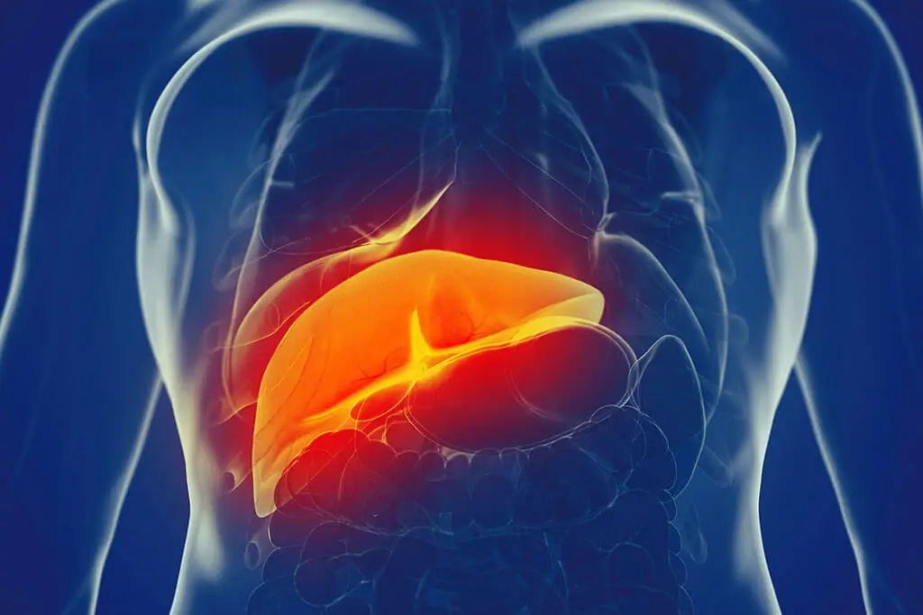
3. Hypoechoic nodule in the liver
Nodules in the liver are associated with a wide variety of conditions. Studies state that in polycystic liver disease, multiple hypoechoic cystic nodules can be found occupying more than 50% of the liver parenchyma.
However, not every hypoechoic lesion is benign and several studies are necessary to rule out malignancy. Nodules with accelerated growth, changes in shape, and a size greater than 1 centimeter (0.4 inches) should be investigated.
What is the treatment for a hypoechoic nodule?
The treatment plan for a hypoechoic nodule varies depending on the organ involved and the morphologic characteristics of the nodule. Most benign lesions require medical supervision every 6 months to monitor their evolution.
On the other hand, nodules that are suspected to be malignant, with changes in their characteristics or accompanied by pain or compressive symptoms, require more aggressive management. Biopsy and puncture for microscopic study is necessary.
In general, malignant lesions should be removed by surgery to avoid possible complications, such as dissemination to neighboring organs.
A rounded hypoechoic nodule that shows low density during ultrasound study may be solid, cystic, or mixed. However, specialist physicians are the only ones qualified to evaluate this type of lesion and provide appropriate treatment.
All cited sources were thoroughly reviewed by our team to ensure their quality, reliability, currency, and validity. The bibliography of this article was considered reliable and of academic or scientific accuracy.
- Díaz-Rodríguez N, Garrido-Chamorro R, Castellano-Alarcón J. Ecografía: principios físicos, ecógrafos y lenguaje ecográfico. SEMERGEN – Medicina de Familia. 2007;33(7):362-369.
- Pavón-Hernández C, Villaseñor-Navarro Y, Cruz-Morales R, Aguilar-Cortázar L, et al. Nódulos, caracterización y categorización. Gaceta Médica de Oncología. 2012; 11(4):260-267.
- López A, Martos J. Pérez M, Pérez I. Nódulo tiroideo. Un viejo problema ante un nuevo siglo. Cirugía Española. 2000; 67(1): 80-93.
- Ramia J, de La Plaza R, Figueras J, García-Parreño J. Tumores hepáticos quísticos benignos no parasitarios. Cirugía Española. 2011;89(9):565-573.
- Gallegos-Hernández JF. Aspectos fundamentales del nódulo tiroideo y el cáncer bien diferenciado de tiroides para los médicos general y familiar. Gac Med Mex. 2019;155(6):619-623.
- Puglia CR. Incidental finding of hepatic nodule. What is its importance?. Rev Assoc Med Bras (1992). 2004 Apr-Jun;50(2):114.
This text is provided for informational purposes only and does not replace consultation with a professional. If in doubt, consult your specialist.


