Hidrocystoma: What It Is, Causes and Treatment

Hidrocystoma is a benign cyst in the sweat glands. It can be eccrine or apocrine, solitary or with many manifestations.
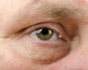
Hidrocystomas contain fluid from the sweat glands trapped inside, without communication with the skin, so there’s no emptying and they can increase in size as more fluid is present. They appear on the skin, predominantly on the edge of the eyelids (especially the lower eyelid), cheeks, and nape of the neck.
They appear more in people living in hot and humid climates.
What causes them?
Hidrocystoma is the result of obstruction of the eccrine sweat gland ducts or regrowth of the apocrine sweat glands. Its exact cause is unknown.
They are associated with hot and humid climates, where more sweat production is required, which could clog the ducts. It also appears more in patients with hyperhidrosis, a condition characterized by excessive sweating.
Regarding hormonal causes, there’s an association with Graves’ disease. In Graves’ disease, there’s hyperstimulation of the sweat glands due to an increase in thyroid hormones.

Hidrocystoma is asymptomatic
The reason for consultation is due to the presence of a cystic nodule that causes aesthetic discomfort. Clinically, hidrocystomas appear as dome-shaped protrusions, usually translucent, but also with pink, bluish, or brownish colorations.
Their surface is smooth and the fluid content is transparent. They can be solitary or multiple.
They can measure between 1 and 16 millimeters and grow or multiply with heat and humidity. When distributed along the eyelid margins they appear lighter in color.
They’re more common in women over 50. Hidrocystomas rarely occur in childhood and adolescence.
The definitive diagnosis is microscopic
The definitive diagnosis is by biopsy or by a precise examination under the microscope. At first glance, hidrocystoma can be confused with other benign lesions, such as hemangiomas, lymphangiomas, dacryocystitis, or a simple dermal cyst.
Malignant lesions also enter the differential diagnosis, such as basal cell carcinoma or melanoma. They’re particularly similar to the former, which is why a biopsy is key to exclusion.
Apocrine versus eccrine hidrocystomas
Eccrine hidrocystomas are smaller and have tighter walls. They predominate in women, on the cheeks and around the eyes.
Apocrine hidrocystomas are usually solitary and larger than apocrine hidrocystomas. They predominate in the head and neck and near the palpebral commissures and palpebral canthus.
Take a look at: Pancreatic Cysts: Causes, Symptoms and Treatment
Treatment
In the case of a solitary hidrocystoma, it’s removed by simple surgery or blepharoplasty surgery. However, the specialist may resort to puncture and draining before attempting this method, because it has a risk of scarring.
In addition, surgical removal may have recurrences.
When there are many of them, topical atropine 1% with scopolamine creams is recommended. Cauterization or electrodesiccation of the wall, laser treatment, or laser vaporization with carbon dioxide can also be used.
The capsule of hidrocystomas is thin, so it can rupture and bleed at the time of exploration or removal. If this happens, it must be ensured that no capsule remains, because they’re conducive to a relapse of the lesion.

Rare lesions that require medical evaluation
Hidrocystomas are rare cystic lesions that represent part of the benign tumors of the sweat glands. They’re small and translucent; cystic ones with fluid content inside may have mild mobilization.
Studying them is important. Due to their similar appearance to other more serious lesions, such as basal cell carcinoma, the appropriate differential diagnostic process is required.
All cited sources were thoroughly reviewed by our team to ensure their quality, reliability, currency, and validity. The bibliography of this article was considered reliable and of academic or scientific accuracy.
- Arceu M, et al. Hidrocistoma apocrino dermatoscópico. Revista Chilena de Dermatología 2019;35(4). Disponible en https://rcderm.org/index.php/rcderm/article/view/260/286.
- Couto A, et al. Hidrocystoma: surgical management of cystic lesions of the eyelid. An Bras Dermatol 2010;85(3). Disponible en https://www.scielo.br/j/abd/a/zCYy49Gj4gzGH8zxT8fXNnD/?format=pdf&lang=en.
- Sarabi K, Khachemoune A. Hidocystomas – A Brief Review. MedGenMed 2006;8(3):57. Disponible en https://www.ncbi.nlm.nih.gov/pmc/articles/PMC1781304/.