Dental CT: Why and How Is It Performed?


Written and verified by the dentist Vanesa Evangelina Buffa
In dentistry, as in other medical specialties, new technologies provide professionals with precise information about their patients. In this article, we’re going to tell you all about dental CT, a cutting-edge tool that provides reliable data on the oral cavity.
With this complementary examination, dentists can make a more accurate diagnosis and plan treatment in detail. Thus, the chances of successful treatment are considerably increased.
Dental CT scanning provides faster, more predictable, and more effective results. Discover what this study of the mouth is all about.
What is a dental CT?
Dental CT is the abbreviated definition of computed axial tomography. It is a device that makes it possible to obtain three-dimensional images of the patient’s teeth and jaws using X-rays.
With this study, it’s possible to obtain images of the dental, nerve, and bone structures of the upper and lower jaws in a single scan. Taking advantage of this information, the dentist can carefully plan treatment before carrying it out.
It’s important not to confuse dental CT with the panoramic X-ray that’s commonly used in dentistry. Although the latter study also provides information on the teeth and bones of the mouth, it doesn’t do so in the three dimensions of a tomography.
A dental CT isn’t routinely used, but only in cases that require a very precise analysis of the craniofacial area. In this sense, it’s a very useful tool in the field of implantology. However, as we will soon tell you, it also has other applications.
The radiation exposure of this type of scanner is greater than in normal dental X-rays. In any case, it’s a safe, simple, and quick test.
We think you may also enjoy reading this article: 9 Natural and Effective Tips for Taking Care of Your Teeth
Why and when is dental CT performed?
Dental CT allows precise three-dimensional images to be obtained of a patient’s entire mouth. With this, the dentist or maxillofacial surgeon can accurately plan the treatment before carrying it out.
However, this is not always necessary. Here are the situations that warrant its indication:
- Implant placement: Dental CT allows the dental surgeon to study the bone structure and determine the exact location and position where the implant is to be placed in the bone tissue.
- Bone regeneration: Before placing an implant, the surgeon must ensure that there’s an adequate quantity and quality of bone to support the screw. The dental CT scan is used to check the patient’s bone dimension and determine if it’s sufficient or if it requires an intervention to increase it. In the event that a sinus lift or grafting is required, the study will provide the necessary data to plan the subsequent intervention. Ultimately, it improves the expected prognosis of the surgery.
- Impacted wisdom teeth: A dental CT helps determine the relationship of impacted molars to other important anatomical structures, such as the inferior dental nerve. It also helps to evaluate the best approach to remove these teeth and reduce adverse effects.
- Included canines: With this study, the position of the canine can be determined, and the best therapeutic alternative can be planned. It’s useful if the tooth is going to be exposed with surgery and repositioned with orthodontics.
- Diagnosis of diseases of the jaws, bone structures of the face, nose, sinuses, and temporomandibular joint disorders (TMJ).
- Detection, measurement, and treatment planning of tumors or cysts in the jaws.
- To locate the origin of pain or a condition that’s difficult to diagnose.
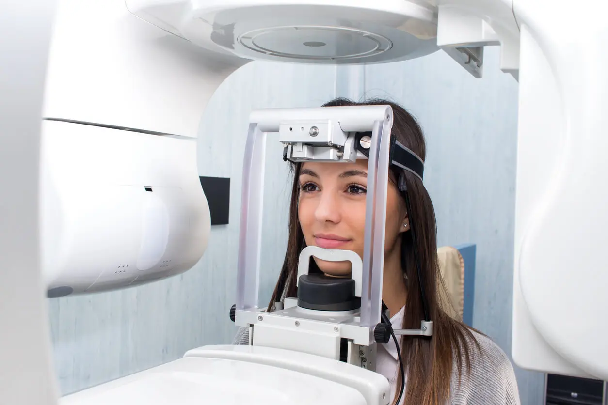
How is a dental CAT scan performed?
The dental CAT scan test is very simple and fast; it only takes a few seconds, and the results can be known instantly. It’s performed in a special X-ray room by qualified personnel. Some dental clinics have this type of technology in their facilities. If this is not the case, the patient should go to an authorized center for these studies and then return to the dentist with the results. The procedure is very similar to that performed for taking panoramic X-rays. No prior preparation is necessary.
At the time of the study, you will need to remove earrings, necklaces, glasses, dentures, hairpins, and any other metal objects. In addition, it will be necessary to protect yourself from radiation by wearing a leaded apron. The professional in charge of performing the dental CT scan places the patient in the exact position so that the apparatus can take the appropriate images of the mouth. This will require the patient to sit in a chair or lie down on the examination table, depending on the type of device being used.
The patient is then positioned so that the area of interest is centered and is asked to remain very still during the study. The X-ray source and detector rotate around the person. In general, the test takes no more than 20 seconds, and during the exam, the patient won’t feel any discomfort or symptoms.
It’s important to remember that pregnant women should not undergo this test. Although the radiation doses used are low, it’s best to avoid these and similar examinations during pregnancy.
Is it dangerous?
A dental CT may be necessary for the diagnosis and planning of various dental treatments. However, since it uses X-radiation, many people wonder if this study carries any health risks.
You should know that the amount of radiation used by this type of scanner is minimal. The radiation of a conventional CT scan is 2 mSv (millisievert). In the case of a digital CT, this figure drops to only 0.02 mSv.
According to the World Health Organization (WHO), the increased risk of cancer or damage to the fetus during gestation associated with the use of radiation occurs in people who receive doses higher than 100 mSv. Therefore, this type of study can be considered safe for our health.

New advances
Medical technology is a field that’s constantly evolving. New advances make increasingly precise equipment available to professionals, offering advantages for both the expert and the patient.
There is, for example, a type of dental CT scanner that’s characterized by the conical shape of its X-ray emission source. This gives the equipment the particularity that, with a single 360-degree rotation, we obtain the necessary information about the area we are interested in. With this, a shorter exposure time is necessary. Therefore, the radiation received by the patient is also lower. This type of cone beam equipment is already in use in several dental centers.
Another state-of-the-art piece of equipment is a new scanner capable of performing panoramic radiography and dental CT at the same time. In general, both studies are needed for an accurate diagnosis. With this test, it’s possible to obtain both in a single exposure.
Like this article? You may also like to read: Seven Home Remedies for Removing Plaque From Your Teeth
A dental CT for successful treatments
A dental CT is a simple, fast, and convenient test. With it, the dentist obtains precise information about the orofacial structures in the three directions of space. Performing this type of study provides data that translate into more accurate approaches. Thus, with a simple test, you increase the chances that the dental procedures indicated to you will be successful and with fewer adverse effects.
All cited sources were thoroughly reviewed by our team to ensure their quality, reliability, currency, and validity. The bibliography of this article was considered reliable and of academic or scientific accuracy.
- Ortega, E. V., & Méndez, Á. G. (2008). La cirugía guiada y carga inmediata en implantología oral. Consideraciones diagnósticas y quirúrgicas. Rev. Esp. Odontoestomatológica de Implantes, 16(4), 211-218.
- COYAC AGUILAR, R. O. G. E. L. I. O. (2010). LA INCLINACION RADICULAR DE CANINOS DETERMINADA CON TOMOGRAFIA, COMPARADA CON SU APRECIACION EN LA RADIOGRAFIA PANORAMICA.
- de-Moraes, L. E., de-Moraes, E. J., Olate, S., & Koch, H. A. (2021). Imagen 3D para la Evaluación Morfológica de Quistes y Tumores Maxilofaciales: Serie de Casos. International Journal of Morphology, 39(4), 1132-1138.
- Edith, G. N. C., Antonio, H. H. J., Frías, M. Á. N., Luciano, A. V. A., Gilberto, L. C., & Rider, R. M. (2014). TAC 3D como estándar de oro para colocación de implantes en pacientes con agenesia dental. Oral, 15(49), 1179-1181.
- ORAL, C. D. E. E. I., & ALTHAPARRO, D. F. C. G. Análisis tomográfico de haz cónico de la frecuencia, ubicación y tamaño de los forámenes linguales para el diagnóstico implantológico en mandíbulas desdentadas.
- Ruiz-Imbert, A. C., & Cascante-Sequeira, D. (2022). Valores de densidad en la escala de grises en Tomografía Computarizada de Haz Cónico: alcances y limitaciones. Odovtos-International Journal of Dental Sciences, 23(2), 52-62.
- Arce Toribio, C. P. Revision bibliografica sobre el uso de acceso guiado por tomografia computarizada en dientes anteriores calcificados.
- Alexandre Oliveira, N., Matos Garrido, N., España López, A., Jiménez Guerra, A., Ortiz García, I., & Velasco Ortega, E. (2019). Planificacion de tratamiento con software para cirugía guiada en implantologia oral. Avances en Odontoestomatología, 35(2), 59-68.
This text is provided for informational purposes only and does not replace consultation with a professional. If in doubt, consult your specialist.


