Alterations in the Position of the Eyelids

Written and verified by the doctor Mariel Mendoza
Palpebral malpositions is the name given to alterations in the correct position of the eyelids, both upper and lower. These abnormal positions can be due to different reasons, although the most common is skin laxity, and then old age.
It is frequently predominant in older adults. In this age group, the prevalence varies from 10% to 17%.
They affect facial harmonization and, in some cases, create vision problems. The symptoms of this alteration in the position of the eyelids depend on each type and five variants have been described:
- Ectropion
- Entropion
- Palpebralptosis
- Dermatochalasis
- Palpebral retraction
The correct position of the eyelids
The eyelids protect the eyeball. They’re responsible for lubrication by blinking.
Tears are produced in the lacrimal glands located above the inner canthus of the eye. This fluid lubricates, cleans, and protects the eyes.
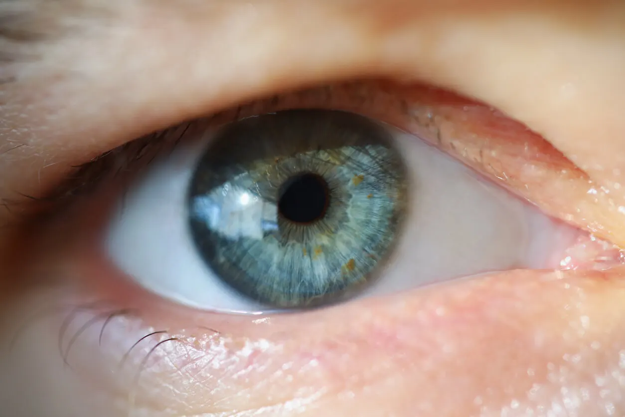
Under normal conditions, the lower eyelid has a free edge that rests on the surface of the eyeball along its entire length. This allows adequate contact with the lacrimal sac and provides the opportunity for the eyelashes to have a forward direction, without impinging on the eyeball.
The free edge of the lower eyelid is held to the middle third of the eyeball, without exposing the sclera or covering the cornea (it’s just between the corneal boundary and the sclera). In turn, the free edge of the upper eyelid covers two millimeters of the cornea.
1. Ectropion
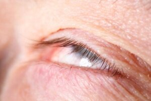
In this condition, the free edge of the lower eyelid is outward, often exposing the eyelid skin and mucosa. Exposure of the mucosa can cause ocular irritation and keratinization.
There is conjunctival redness, foreign body sensation, excessive tearing (epiphora) and it predisposes to infections on the surface of the eye. Its main cause is the loss of tissue elasticity (skin laxity) due to aging.
It can also originate in facial nerve paralysis, scars from trauma, or previous surgeries. It may be a consequence of artificial tears used for glaucoma, excessive sun exposure, and ocular tumors.
The initial treatment is the use of artificial tears, but its definitive correction is surgical.
2. Entropion
In these palpebral malpositions, the free edge of the lower eyelid turns inward. Thus, the eyelid skin and eyelashes come in direct contact with the cornea and can be returned to their original position only when the eye is tightly closed. The contact of the eyelashes with the cornea can generate from intraocular infections to corneal abrasions or ulcers.
Its causes are varied and include the following:
- Skin diseases leading to laxity
- Skin weakness due to age
- Trauma that has caused scarring
- Facial nerve paralysis
- Muscle weakness
- Corneal defects
- Previous surgery
- Ocular infection
Symptoms are similar to ectropion (epiphora, blurred vision, red eye, foreign body sensation), but it’s associated to a greater extent with visual field reduction and it’s more aggressive because it has direct contact with the cornea. It’s treated surgically.
In some cases, it can be relieved with special eye drops. Botox injections are also used.
3. Palpebral ptosis
Palpebral ptosis refers to the lowering or drooping of the upper eyelid below its normal position. It’s defined by an asymmetry of more than 2 millimeters between the opening of both eyes or a descent of more than 2 millimeters of the upper eyelid over the eyeball.
From the clinical point of view, in this type of palpebral malposition, the main symptom is the impossibility to maintain an adequate opening of the eye. Consequently, the visual field is also affected.
Its origin can be congenital or acquired. In the former, we’re talking about lax eyelid syndrome. The acquired one may be due to skin laxity, eyelid edema, tumors in the orbit, or problems with the ocular musculature, such as myasthenia gravis or essential blepharospasm.
Treatment is only surgical and requires intervention in the ocular muscles.
Learn more about: Ptosis or Droopy Eyelid: Causes and Treatments
4. Dermatochalasis
Among the different problems affecting the position of the eyelids, we have dermatochalasis. It’s a redundancy and laxity in the eyelid skin, predominantly in the upper eyelid, with an accumulation of wrinkles and fatty tissue bags.
It manifests as a bulging of the upper eyelid or the presence of multiple small bags in the lower eyelid. There may be alterations in the visual field due to obstruction.
Most sufferers are older (over 60 years of age). However, there may also be a genetic component, as occurs in the flaccid eyelid.
Its resolution is surgical, by means of blepharoplasty. The fat bags are resected and the excess tissue is removed.
Read about: 7 Tips for Treating Eye Tic Disorder
5. Palpebral retraction
This is simply an abnormal high position of the upper eyelid or low position of the lower eyelid. Thus, the eyeball isn’t properly covered and exposed to external agents.
The clinical manifestations will depend on which agent contacts the eye. In addition to the aesthetic alteration in the harmonization of the face. There are usually recurrent eye infections and blurred vision.
Its causes are varied and include the following:
- Tumors (teratomas)
- High grade myopia
- Thyroid disease
- Tissue laxity
- Affection in the size of the orbital bones
Treatment is only surgical.
Palpebral malpositions and surgery
Because the causes are so varied and predominantly associated with aging, palpebral malpositions can’t be prevented, but they can be treated early. That’s why it’s important, in case of any suspicious symptoms, to go and see an ophthalmologist.
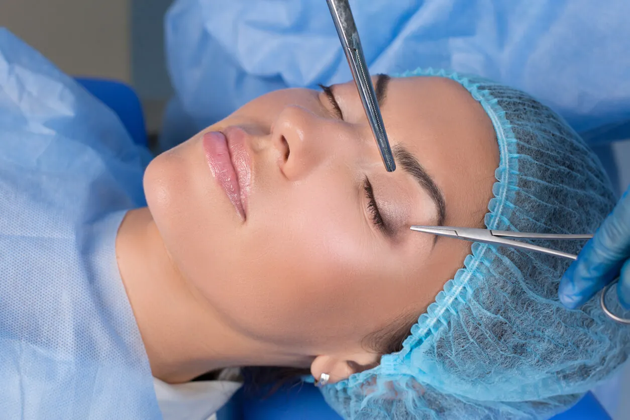
Its diagnosis is simple, and by means of an ophthalmologic examination. Its definitive treatment is surgical.
It’s carried out with repositioning surgery or anatomical and functional reconstruction of the eyelid. It can be performed externally (through the skin) or internally (through the conjunctiva). This surgery doesn’t require hospitalization and gives few complications.
All cited sources were thoroughly reviewed by our team to ensure their quality, reliability, currency, and validity. The bibliography of this article was considered reliable and of academic or scientific accuracy.
- An, S. H., Jin, S. W., Kwon, Y. H., Ryu, W. Y., Jeong, W. J., & Ahn, H. B. (2016). Effects of upper lid blepharoplasty on visual quality in patients with lash ptosis and dermatochalasis. International Journal of Ophthalmology, 9(9), 1320.
- Batista Peña, M., Rojas Rondón, I., Arzuaga Hernández, E., & Guzmán Martínez, M. D. L. (2019). Síndrome de párpado laxo. Revista Cubana de Oftalmología, 32(4).
- Bergstrom, R., & Czyz, C. N. (2022). Entropion eyelid reconstruction. In StatPearls [Internet]. StatPearls Publishing.
- Hakim, F., & Phelps, P. O. (2020). Entropion and ectropion. Disease-a-Month, 66(10), 101039.
- Pellerano, F., Guillermo, E., Garrido, G., & Berges, P. (2017). Congenital orbital teratoma. Ocular oncology and pathology, 3(1), 11-16.
This text is provided for informational purposes only and does not replace consultation with a professional. If in doubt, consult your specialist.


