What's a Craniotomy and When's It Necessary?

Multiple conditions can affect brain functions and performing a craniotomy is often the only possible solution. It’s an important one as the brain is the control center of all body functions.
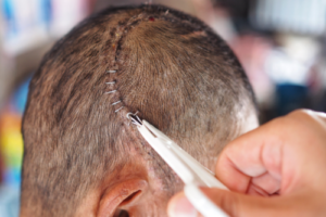
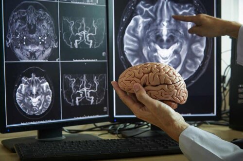
A craniotomy is a surgical procedure that consists of the removal of a segment of the skull to expose part of the brain. The removal of the bone is temporary and the surgeon will put it back in place before they finish the surgery.
Of course, it must be done by a neurosurgeon who’ll determine the size of the incision, depending on the problem. One of the major uses of this procedure is the removal of brain tumors.
What’s the purpose of a craniotomy?
This procedure is appropriate for patients with structural and other types of alterations that affect proper brain functioning. Its main objective is to repair any existing abnormality without affecting the cortical functions.
Thus, a craniotomy provides access to the brain to remove or repair something. Furthermore, it’s a rather complex and delicate procedure and some people dread it. Nevertheless, its success rates are quite high.
Read about The Risks of a Sedentary Lifestyle for Your Brain
Craniotomy versus craniectomy
People often mistake “craniotomy” with another similar procedure called “craniectomy.” However, this latter one is the permanent removal of a segment of the skull. That is doctors don’t put back the removed bone in place at the end of it.
The main function of a craniectomy is to provide more space for the brain when there’s a significant inflammatory process such as intracranial hypertension. This alteration has multiple causes, such as hemorrhages, infarctions, and cranioencephalic trauma.

Conditions that require a craniotomy
One of the main reasons for performing this surgery is the presence of a brain tumor. In these cases, studies show that leaving certain brain nuclei active during the procedure helps preserve all major functions. A craniotomy may also be the solution for the following conditions:
- Aneurysms or dilation of cerebral arteries
- Subdural, epidural, and intracerebral hematomas
- Brain abscesses
- Congenital arteriovenous abnormalities
- Lesions in the meninges
- Cranial fractures following trauma
Before surgery
The preparation for the procedure is quite rigorous as it’s quite invasive. First, the physician must order all sorts of lab tests to find out if they could perform it successfully.
People must usually wash their hair with a special antiseptic shampoo the night before the craniotomy. This is to reduce the risk of infection. In addition, they must skip any slow-digesting foods on the day of surgery. In some cases, doctors also recommend shaving the entire head or at least cutting the hair really short.
Specialists may indicate other previous measures, depending on the underlying pathology. For instance, a person may have to suspend the consumption of certain drugs up to a week before the procedure. They must also avoid drinking alcohol and smoking.
Once in the hospital, the patient and their family must sign a series of documents and a nurse will explain the whole procedure. In addition, the anesthesiologist will provide information about general anesthesia and its possible side effects.
What’s the procedure like?
In general terms, this kind of operation can last between three and five hours, although everything will depend on the condition under treatment.
Once inside the operating room, the patient will lie down on the operating table and the medical staff will install an intravenous line, a bladder catheter to empty the bladder, and general anesthesia.
After the anesthesia begins to do its thing, the health personnel will proceed to remove all the hair from the incision site and clean the area. The doctor will then cut the scalp with a scalpel and drill small holes with a specialized drill. Finally, they’ll cut the desired segment of bone with a craniotome tool.
The tough outer meninx, called the dura, will be visible and will need to be cut to expose the brain after removing the bone. The doctor will then use a series of small instruments to move around inside. They’ll also need lenses with special magnification.
After repairing the brain abnormality, the specialist must carefully remove all instruments and suture the dura. Then, they’ll place back the extracted bone segment in its original position, fixing it to the skull with titanium plates and screws.
Have you heard about The Brain-Eating Amoeba: Naegleria Fowleri?
The recovery process after a craniotomy
People undergoing surgery usually go into a recovery room and then the medical staff transfers them to the intensive care unit (ICU) after they wake up and seem stable.
Health care providers must make sure there are no further brain conditions. All people who undergo this surgery usually have a sore throat and nausea during the first few days and these can go away with medication.
Multiple studies show that headaches are also frequent after the surgery, especially during the first days. This headache appears on the same side of the procedure but improves after a few weeks. Opioids, anti-inflammatory drugs, and analgesics can mitigate the discomfort.
Hospital stay varies from two days to two weeks. In addition, patients must attend a follow-up medical consultation two weeks after the surgery. The total recovery time varies, it ranges from one to four weeks.
Possible risks and complications
A craniotomy is a rather delicate operation and so complications may occur. First of all, there are those associated with all types of surgeries, such as reactions to medications, respiratory problems, and hemorrhages.
In addition, this surgery has particular risks and complications, the most frequent being alterations in brain function. In this respect, people may experience alterations in speech, memory, and coordination.
All these are due to possible brain and nerve damage, generally during the removal of a tumor. Other frequent complications are:
- Muscle weakness
- Convulsions
- Cerebral vascular disease (CVD)
- Coma
- Bleeding, infection, or cerebral edema

Lifestyle and recommendations
Outcomes and prognosis following a craniotomy vary. However, they’re favorable in most cases. The surgery can affect a person’s lifestyle, especially during the first few weeks afterward.
In this regard, it’s important to maintain adequate rest, limit exertion, and perform appropriate physical therapy sessions until fully recovering. Everyone should improve their lifestyle and adopt healthier habits though.
A procedure with a high success rate
A craniotomy provides access to the encephalic mass, which allows solving multiple brain conditions. It’s the best solution for aneurysms and multiple tumors, since it allows repairing the alteration while generating the least possible damage.
This procedure has a large number of possible complications, but the benefits outweigh the risks. As always, it’s important to inform the doctor about any questions or concerns.
All cited sources were thoroughly reviewed by our team to ensure their quality, reliability, currency, and validity. The bibliography of this article was considered reliable and of academic or scientific accuracy.
- Kobyakov GL, Lubnin AY, Kulikov AS, Gavrilov AG et al. Awake craniotomy. Zh Vopr Neirokhir Im N N Burdenko. 2016;80(1):107-116.
- Rocha-Filho PA. Post-craniotomy headache: a clinical view with a focus on the persistent form. Headache. 2015 May;55(5):733-8.
- González-Darder JM. Historia de la craneotomía [History of the craniotomy]. Neurocirugia (Astur). 2016 Sep-Oct;27(5):245-57.
- Shimizu T, Aihara M, Yamaguchi R, Sato K et al. Large Craniotomy Increases the Risk of Minor Perioperative Complications in Revascularization Surgery for Moyamoya Disease. World Neurosurg. 2020 Sep;141:e498-e507.
- Morales-Valero SF, Van Gompel JJ, Loumiotis I, Lanzino G. Craniotomy for anterior cranial fossa meningiomas: historical overview. Neurosurg Focus. 2014 Apr;36(4):E14.
- Kennion O, Holliman D. Outcome after craniotomy for recurrent cranial metastases. Br J Neurosurg. 2017 Jun;31(3):369-373.
This text is provided for informational purposes only and does not replace consultation with a professional. If in doubt, consult your specialist.


