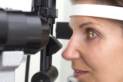
Description and Characteristics of Retinitis Pigmentosa


Written and verified by the doctor Leonardo Biolatto
The disease known as retinitis or retinitis pigmentosa is actually a group of pathologies that have in common a genetic alteration that modifies the reception of light in the retina. The cells of the fundus of the eye progressively degenerate as the pathology evolves.
It’s a rare disorder with an incidence of 1 out of 3,000 people. In any case, it’s the most common of this group if you consider all the dystrophies transmitted from parents to children. What are the causes? Continue reading to find out.
Causes of retinitis pigmentosa
This disease is genetic and hereditary. What this means is that parents pass it on to their children due to a gene mutation. We say it’s a group of disorders, and not just one, because researchers already identified thousands of changes in DNA (deoxyribonucleic acid) that culminate in retinitis pigmentosa.
Even so, it’s impossible to find the origin of the disease in about a third of those afflicted by it. No doubt it’s genetic, it’s only that it is impossible to reach the mutation that degenerates the cells in these people.
Its inheritance has been classified into three groups due to this variability:
- By the X chromosome. In these cases, only men have symptoms while women are carriers and don’t ever develop the pathology. Unfortunately, they do pass it on to their male children.
- Dominant transmission. This means a given gene always expresses. In this type of retinitis pigmentosa, all generations of the same family with the mutation end up losing their vision.
- Recessive transmission. Unlike the previous one, this gene doesn’t always manifest so a parent might have symptoms but their children don’t, and yet their grandchildren do.

Keep reading about the Ten Exercises and Foods for Good Vision
Symptoms of the disorder
This disease coincides with some symptoms that are general for all patients — despite responding to different mutations. The first signs usually appear during adolescence.
The progression of retinitis pigmentosa gradually leads to the loss of vision. However, it isn’t always the same type of loss. The most frequent forms are:
- Color confusion. This is similar to color blindness and patients mistake one color for another or don’t identify it as such.
- Loss of central vision. This is the vision we use most for tasks that require concentration in a short distance. In fact, reading becomes difficult if the patient loses this ability. This is because it has to do with the area of the retina that suffers from the disorder in the first place. Consultation for this symptom is often confused with hyperopia — especially if the person is over 40 years old.
- Tunnel vision. This is the opposite of the previous process. Here, the patient loses the ability to distinguish shapes and colors in the periphery of their visual field. Thus, they can only see what’s in the center and very close. The name “tunnel” has to do with the sensation experienced by those who suffer from it. It isn’t unique to retinitis pigmentosa and, therefore, is often confused with other pathologies.
- Decreased ability to see in the dark. This is one of the most frequent manifestations and thus the one that most guides the diagnoses. It means the patient can see normally during the day but tends to be nearly blind when it gets dark or when the light is turned off in a given environment.
Diagnosis
The first thing a doctor considers is the suspicion of retinitis pigmentosa. They’ll have a hypothesis from the beginning if a person has a family history of it. However, the task becomes more difficult if there are no previous records between parents or grandparents.
The physician will order a genetic test once they establish the symptoms. They analyze the composition of the genes to locate those that are defective and that could explain the disorder through a blood sample.
Furthermore, they may complement the evaluation with electroretinography, the measurement of the electrical activity of the retina, and also with an optical coherence tomography. The latter is a series of images obtained from the retinal tissue in high definition.
The visual field is a test that’s always performed in ophthalmology and doesn’t contribute to the diagnosis. It’s useful to establish the degree of involvement of a given patient. This is because it’s possible to know how much the progression has evolved based on the remaining visual capacity.

Find out more about How to Use Pressed Garlic to Help With Vision Loss
Treatment of retinitis pigmentosa
There’s no cure for retinitis pigmentosa. The treatments are mainly about trying to reduce the symptoms and slow down the progression of blindness to improve the quality of life of an affected person.
There’s been successful experimentation with high doses of vitamin A in the chemical form of palmitate. However, it’s neither clear if this is a treatment of choice, nor if the adverse effects of these concentrations aren’t a risk to other organs such as the liver.
Doctors also prescribe anti-inflammatory drops if there are bothersome symptoms such as itching, pain, gritty eyes, or redness. These drugs are for relief only but aren’t chronic management options in any way.
In addition, there’s the option of surgery in specific cases such as if the disease is accompanied by cataracts. In addition, the artificial retina implant is still under study to determine how effective it is.
The importance of prenatal consultation
Finally, because this is a hereditary disease, prenatal consultation in retinitis pigmentosa is essential. Anyone who’s considering having children and has a diagnosis of the pathology should know about it. There’s no way to avoid it, but they may be able to take some precautions.
Consult your doctor if you present any ocular symptoms or notice your vision is failing. An ophthalmologist should know how to perform complementary methods to address the problem.
We hope you’ve enjoyed this article.
All cited sources were thoroughly reviewed by our team to ensure their quality, reliability, currency, and validity. The bibliography of this article was considered reliable and of academic or scientific accuracy.
- Triana Casado, Idalia, et al. “Caracterización clínicoepidemiológica de pacientes con retinosis pigmentaria y glaucoma.” Medisan 15.5 (2011): 604-610.
- Hernández Baguer, Raisa, et al. “Retinosis Pigmentaria: clínica, genética y epidemiología en estudio de familias habaneras.” Revista Habanera de Ciencias Médicas 7.1 (2008): 0-0.
-
Musarella MA. Mapping of the X-linked recessive retinitis pigmentosa gene. A review. Ophthalmic Paediatr Genet. 1990;11(2):77-88. doi:10.3109/13816819009012951
- Pérez Díaz, Maritza, et al. “Retinosis Pigmentaria en niños y adolescentes: características psicológicas.” Rev. cuba. oftalmol 6.1 (1993): 25-33.
- Ferrari S, Di Iorio E, Barbaro V, Ponzin D, Sorrentino FS, Parmeggiani F. Retinitis pigmentosa: genes and disease mechanisms. Curr Genomics. 2011;12(4):238-249. doi:10.2174/138920211795860107
- de la Mata Pérez, G., et al. “Retinosis pigmentaria sine pigmento. Inicio con edema macular.” Archivos de la Sociedad Española de Oftalmología 89.9 (2014): 376-381.
- Delgado-Pelayo, Sarai A. “Retinosis pigmentaria.” Revista Médica MD 3.3 (2012): 163-167.
- Dyce Gordon, Elisa Idalmi. “Aspectos genético-sociales de la retinosis pigmentaria.” Revista Archivo Médico de Camagüey 14.2 (2010): 0-0.
- Castaño, Alberto Jesús Barrientos, et al. “Fenotipo electrorretinográfico en pacientes con Retinosis Pigmentaria.” Revista Cubana de Genética Comunitaria 6.1 (2012): 20-25.
- Achar, Víctor, and Juan Carlos Zenteno. “Actualidades en el tratamiento de la retinosis pigmentaria.” Revista Mexicana de Oftalmología 82.5 (2008): 309-313.
- Benito Pascual, Ana María. “Restauración de la función visual con el implante de retina Argus II.” (2017).
This text is provided for informational purposes only and does not replace consultation with a professional. If in doubt, consult your specialist.


