Colored Marks on Eyelids: Why Do They Appear?

Written and verified by the dermatologist Maria del Carmen Hernandez
The colored marks that can appear on eyelids have multiple causes, from metabolic illnesses to allergic reactions. Therefore, they tend to differ in color and thickness. As such, they can be representative of several factors.
In general, their presence relates to hormonal alterations, use of medication, excessive exposure to ultraviolet rays, among other factors.
What are their characteristics? What treatments are available to treat them?
Keep reading to find out!
Colored marks on eyelids
Not all of the colored marks that appear on eyelids are related to poor sleep, and the bags that form underneath. Some of them are indications of an illness that requires a particular treatment.
In this article, we’ll explain what some of them are.
Xanthelasma
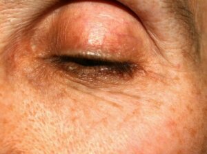
Xanthelasma is yellow-ish papules and plaques caused by an accumulation of lipid deposits. They usually form on the eyelids and can indicate a plasma-lipid disorder, as seen in approximately 50% of people.
Therefore, they must be evaluated to detect if the underlying cause is hyperlipidemia. However, it’s an asymptomatic benign lesion, as it isn’t related to complications affecting the skin.
That said, some people prefer to treat them for cosmetic reasons. Of these, surgical removal is one of the most commonly used procedures due to its aesthetic results.
Hormonal changes during pregnancy
The hormonal changes that the body produces during the gestation period are related to changes in the skin. In particular, it usually causes a condition known as melasma. Melasma causes hyperpigmented tinges and symmetrically distributed patches on the face, neck, and in some cases the upper part of the body.
The known risk factors include ultraviolet radiation (UV), hormonal variations, thyroid diseases, and the use of contraceptives. In the case of those who have marks on their eyelids due to this condition, the treatment options are as follows:
- Topical hyper-pigmenting agents
- Chemical peels
- Laser therapy
- Superficial microdermabrasion
You may also be interested in: Three Natural Remedies for Melasma
Prostaglandin analogs for glaucoma
The prostaglandin analogs that are used to treat glaucoma, like latanoprost and bimatoprost, can cause dark eyelids after approximately 3 to 6 months of use.
A publication in the American Journal of Dermatology concludes that when people stop using bimatoprost, the dark circles can disappear completely after some time.
Dermal melanocytosis
Dermal melanocytosis is an umbrella term for a series of skin lesions that are characterized by the presence of marks. In particular, those suffering from this condition have grey or greyish-blue marks on their eyelids. This can be due to acquired or congenital causes.
The articles in the Journal of the American Academy of Dermatology explain that their presence is related to sun exposure, hormonal changes during pregnancy, and chronic atopic dermatitis.
Vitiligo
Vitiligo is the selective destruction of melanocytes. As a result, patches of depigmentation appear on the skin. Vitiligo of the eyelids, eyebrows, and genitals are a therapeutic dilemma, especially in children.
There’s a strong link between those with vitiligo and those with eye disorders, according to studies published in the Journal of the Indian Medical Association. In these cases, the immunomodulating agent pimecrolimus could be a suitable treatment option.
Post-inflammatory hyperpigmentation
Chronic eczema and inflammation of the eyes, due to allergies, can cause hyperpigmentation and dark eyelids. Some people may begin to rub and scratch the skin around this zone. As such, it can also be the result of a build-up of fluid due to the allergies.
Also read: The Causes of Hyperpigmentation
Amyloidosis purpura
Amyloidosis is an uncommon disorder that occurs when a protein called ‘amyloid’ is found in the organs. It’s an illness that doesn’t have a cure, but some treatments contribute to controlling its symptoms and limiting the production of the amyloid substance.
The appearance of atraumatic purple marks on the face – mainly eyelids and neck – is probably an indicator of amyloidosis. To receive a suitable diagnosis, you should visit a medical professional.
Orange palpebral spots (OPS)
Orange palpebral spots manifest on the internal part of the upper lid, which differs from xanthelasma. It isn’t completely clear where it comes from, but professionals believe it generates from lipofuscin deposits.
Treat the colored marks on your eyelids for aesthetic reasons
Having colored marks on your eyelids is a common condition and generally doesn’t represent anything serious. However, those that suffer often look for treatment options as they consider them unsightly.
Fortunately, there are currently several creams and aesthetic procedures that help to minimize them to improve the overall appearance of the skin. It’s worth consulting a dermatologist to choose the best option according to your specific case.
All cited sources were thoroughly reviewed by our team to ensure their quality, reliability, currency, and validity. The bibliography of this article was considered reliable and of academic or scientific accuracy.
- Hori Y, Kawashima M, Oohara K, Kukita A. Acquired, bilateral nevus of Ota-like macules. J Am Acad Dermatol. 1984;10(6):961-964. doi:10.1016/s0190-9622(84)80313-8
- Sodhi PK, Verma L, Ratan SK. Increased periocular pigmentation with ocular hypotensive lipid use in African Americans. Am J Ophthalmol. 2004;137(4):783. doi:10.1016/j.ajo.2003.11.073
- Ing EB, Buncic JR, Weiser BA, de Nanassy J, Boxall L. Periorbital hyperpigmentation and erythema dyschromicum perstans. Can J Ophthalmol. 1992;27(7):353-355.
- Cho S, Lee SJ, Chung WS, Kang JM, Kim YK, Cho SB. Acquired bilateral nevus of Ota-like macules mimicking dark circles under the eyes. J Cosmet Laser Ther. 2010;12(3):143-144. doi:10.3109/14764172.2010.487912
- Lee HY, Jin US, Minn KW, Park YO. Outcomes of surgical management of xanthelasma palpebrarum. Arch Plast Surg. 2013;40(4):380-386. doi:10.5999/aps.2013.40.4.380
- Biswas G, Barbhuiya JN, Biswas MC, Islam MN, Dutta S. Clinical pattern of ocular manifestations in vitiligo. J Indian Med Assoc. 2003;101(8):478-480.
- Assouly P, Cavelier-Balloy B, Dupré T. Orange palpebral spots. Dermatology. 2008;216(2):166-170. doi:10.1159/000111516
This text is provided for informational purposes only and does not replace consultation with a professional. If in doubt, consult your specialist.


