Chondromalacia Patellae - Characteristics and Treatment
Chondromalacia patellae are one of the most common knee conditions. Keep in mind this joint is highly prone to injury as it supports most of the human body weight. Additionally, there are other risk factors such as age, overweight, level of activity, etc.
Note that chondromalacia patellae are a relatively common condition among young adults. There’s a higher incidence among those who practice soccer, basketball, volleyball, tennis, cycling, karate, rowing, rugby, athletics, and ballet. Mountaineers are also highly prone to it.
There’s a debate around the use of the term chondromalacia patellae. The first word refers to the softening of the cartilage of the knee. However, it began to be used to diagnose anyone with pain in that joint.
Currently, the scientific community thinks it’s more accurate to speak of patellofemoral syndrome and not of chondromalacia patellae when the exact cause of knee pain is unknown. However, the term is medically acceptable.
What are chondromalacia patellae?
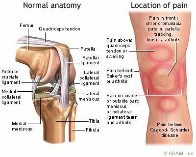
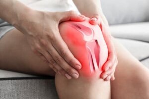
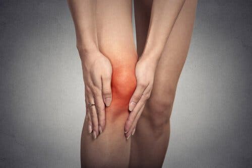
The patellae are flat bones located on the anterior side of the knee. They articulate the femur and their main function is to protect and facilitate the sliding of the joint. Chondromalacia patellae is a degenerative condition that affects the cartilage located on the articular surface of the patellae.
When this condition occurs, the bone is exposed to direct rubbing, due to joint movement. In most cases, this is due to the softening of the cartilage, causing pain on the anterior side of the knee.
The cartilage acts as a shock absorber in the joints. It’s made up of 90% water and 10% cells. This allows it to withstand the energy of the impacts. When there’s chondromalacia patella it’s because the cartilage changed its structure. Thus, it stops being smooth and becomes rough and gray.
The cartilage then becomes thinner and more irregular. Sometimes it has cracks and even breaks. In those cases, the knee makes some clicks and sounds when doing some movements. There’s also severe pain.
Discover Home Remedies to Treat Runner’s Knee
Causes of the problem
The main cause of chondromalacia patella is repeated trauma to the knee. In other words, repeated movements generate compression in the cartilage. This is why athletes are more exposed to this condition.
Likewise, the problem may be due to other factors such as:
- Firstly, anatomic problems of knee misalignment or poor kneecap placement
- Also, strong blows to the knee when someone falls on their flexed knee or impacts it against an object
- Inadequate running that stems from an abnormality in the feet or legs, or from wearing high heels
- A previous knee dislocation or a fracture
- Being overweight
- Muscular atrophy or weakness
- A disparity in the length of the legs
- The prominent curve in the spine
You may be interested: Learn All About Kneecap Dislocation
Characteristics
Typically, there’s a pain when doing physical activity. It increases when the patient runs on hard surfaces, or when going up and down the stairs. Also when they stay in a position that requires maintaining your knees bent for a long time, in which case you also experience stiffness.
When flexing the knee, it’s common for the characteristic clicks of chondromalacia patellae to occur. Over time, this abnormality affects the strength of the leg muscles and walking.
According to the evolution of this condition, there are various levels of severity. These are:
- Grade 1. The cartilage softens and there is edema
- Grade 2. Diagnostic images reveal that the cartilage has fibrillation, there’s fraying
- Grade 3. The cartilage presents fissures, some of which are pronounced and reach the deepest layers
- Grade 4. There’s ulceration. Grade 3 condition aggravates and is more evident
- Finally, there’s grade 5. Here, there’s eburnation, that is, a pathological increase in cartilage density — an effect of worsening ulceration, which ends up affecting the subchondral bone.
The most common conservative treatment is first applied to manage this condition. If this doesn’t produce results, then it’s necessary to undergo surgery.
https://mejorconsalud.com/que-es-condromalacia-rotula/
All cited sources were thoroughly reviewed by our team to ensure their quality, reliability, currency, and validity. The bibliography of this article was considered reliable and of academic or scientific accuracy.
Sánchez, K. T. R. (2014). Condromalacia rotuliana. Revista Médica de Costa Rica y Centroamérica, 71(611), 551-553.
This text is provided for informational purposes only and does not replace consultation with a professional. If in doubt, consult your specialist.


