Surgical Removal of Exostoses of the External Auditory Canal
Surgical removal of exostoses of the external auditory canal is indicated in cases where there’s a total, or almost total, blockage of the external auditory canal.
In the initial stages, as well as in the more advanced ones, ear cleaning is very difficult and, sometimes, almost impossible. This will make external otitis very recurrent. In fact, it may even cause hearing loss.
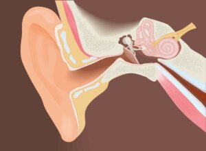
Exostoses of the external auditory canal
Exostoses of the external auditory canal are benign bone formations. These bone growths occur in the ear canal, causing it to narrow. They’re generally bilateral formations. They manifest on the anteroinferior and posterior walls of the ear’s external auditory canal.
Although the exact cause is unknown, the strongest theory is that irritations that originate in the external duct from repeated contact with cold water cause them. For this reason, exostoses of the external auditory canal are more common in people who practice water sports.
Does it cause symptoms?
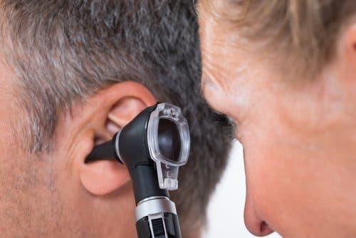
Most of the time, exostoses don’t cause symptoms. They’re casually diagnosed during an ear check. In general, they evolve very slowly and produce no symptoms at first.
However, when they grow and close the duct, the first discomfort begins to appear. The growth of exostoses favors the retention of earwax and water in the ear canal. As a consequence of this retention, recurring external otitis will occur.
Generally, if the obstruction of the external auditory canal is less than 60%, there are usually no problems. However, in patients with an obstruction greater than 80%, there’s a higher incidence of external otitis and sensorineural hearing loss.
Read on to learn more: Hearing Loss: Symptoms and Treatment
Treatment
To treat milder cases of exostoses of the external auditory canal, ear vacuuming and a general cleaning of the ear is enough. In addition, it’s a good idea to do follow-up controls to observe the growth. This way, you can avoid complications in the early stages of the disease. However, for infections, medical professionals often prescribe the use of antibiotic ear drops.
Surgical removal of exostoses of the external auditory canal

In the most severe cases of exostoses, when the ear canal stenosis is practically complete, surgery is used to remove it.
There are various surgical techniques of removal of exostoses of the external auditory canal. Some of them are with local anesthesia, while others need general anesthetic. Even the duct approach can be retroauricular or endaural.
Also, there are many different methods to eliminate exostoses. In this regard, some techniques carry out milling, others use a chisel, and others laser. In addition, some surgical techniques remove them from all the walls of the external auditory canal, others only from the anterior wall, and others only from the posterior wall.
Possible complications of the surgical removal of exostoses
The surgical removal of exostoses isn’t easy, and numerous complications can derive from it. Some of the most common complications are:
- Postoperative canal stenosis
- Facial nerve injury
- Eardrum perforation
- Injuries to the temporomandibular joint
- Sensorineural hearing loss
Doctors surgically remove exostoses under general or local anesthesia and through an incision behind the ear and under a microscopic view. The patient is normally discharged after an overnight stay. The doctor removes the stitches after one week and the earplug after two weeks.
The patient can resume their work-life in 7-14 days. Doctors generally recommend not wetting the ear until the new duct heals properly. This healing process can take between one and three months. Regarding post-surgical results, in most cases, it solves the problems of blockage and otitis.
This article may also interest you: Protect Your Hearing: When Was Your Last Checkup?
Can they be prevented?

While it’s true that many water sports athletes use earplugs, their use hasn’t been shown to be effective in preventing exostoses.
Even sometimes, in patients who already have exostoses, the plugs can push the wax in and worsen the condition. The specialist will assess each case and, depending on the patient’s habits and the degree of exostoses, will recommend the best option.
All cited sources were thoroughly reviewed by our team to ensure their quality, reliability, currency, and validity. The bibliography of this article was considered reliable and of academic or scientific accuracy.
- Granell, J., Puig, A., & Benito, E. (2013). Osteoma y exóstosis del conducto auditivo externo: un diagnóstico clínico. Acta Otorrinolaringológica Española. https://doi.org/10.1016/s0001-6519(03)78408-8
- Rivera Rodríguez, T., & Olarieta Soto, J. (2013). El paciente con hipoacusia. Medicine – Programa de Formación Médica Continuada Acreditado. https://doi.org/10.1016/s0304-5412(01)70552-5
- Bejarano-Panadés, N., Corral-Juan, J. L., & Juan-Fernández, J. M. (2007). Enfermedades del oído externo y la articulación temporomandibular en el buceo. Acta Otorrinolaringol Esp.
This text is provided for informational purposes only and does not replace consultation with a professional. If in doubt, consult your specialist.


