How Coronavirus Infects Cells: The Latest Stunning Images
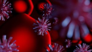
The National Institute of Allergy and Infectious Diseases (NIAID) recently released electron microscopy images of how the coronavirus infects cells. Click here to see these incredible images which are available on the institution’s Flickr account.
Under electron microscopy, these small worlds that are invisible to the human eye can be photographed. Then, through an editing process, colors are added and removed to highlight what has been observed. In this case, NIAID used a state-of-the-art electron microscope from the Integrated Research Facility in Maryland. There, they analyzed samples from a SARS-CoV-2 infected patient.
These images provide an overview of the infectious process of the virus that’s causing the current global pandemic. The pandemic began in late 2019 in Wuhan, China, and continues to spread throughout the planet, and has infected more than 500,000 people at this time.
How the coronavirus infects cells
Knowing how the coronavirus infects cells is key to preventing and treating the disease. As many have said, we’re facing an invisible enemy that we know very little about because of its recent appearance.
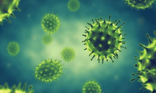
The coronavirus responsible for this pandemic is the SARS-CoV-2 variety. Scientists assume that it’s a mutation of a coronavirus in an animal that then acquired the ability to perform inter-human transmission.
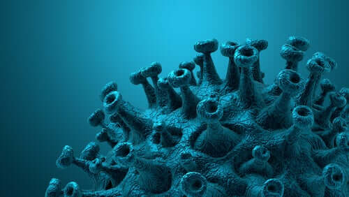
The coronavirus in question is no more than 160 nanometers in diameter. This is a tiny measurement that’s impossible to imagine. It’s even smaller than the cells of the human body and needs to penetrate them to use their structure. It’s an RNA virus, which means that its genetic information is stored in ribonucleic acid. To replicate and multiply, the coronavirus uses its host’s cells. There, it makes new RNA and proteins to cover itself and then continue on its way.
The coronavirus infects cells by penetrating them with its spicule covering. The prefix “corona” in its name refers to the spikes that surround it in a crown form. That’s where it houses a key protein that contains the SARS-CoV-2 virus prepared for entry.
Which cells are most affected by the coronavirus?
The human cells most affected by the coronavirus are cells in our lungs. That’s where the main symptoms come from. Coughing is the result of an irritation of the airways, and pneumonia is the complication that can lead to death.
The spicules of the SARS-CoV-2 coating attach to the receptors on the pulmonary alveoli. They do this like a key opening a lock, and, in this way, infect the cells with coronavirus. Once inside, they replicate, creating new particles.
The infected lung then starts to fill with immune system cells. White blood cells arrive at the area of infection to try to fight the foreign agent. Once there, they release immune substances and antibodies that mix with the inflammatory fluid. The fluid that collects in the lung tissue is what you can see on x-rays as a type of frosted glass effect. The problem with this liquid is that it isn’t absorbed and becomes fibrous tissue, like a large scar.
Coronavirus-infected lung cells don’t function normally. Because the lung contains the tissue that’s responsible for breathing, this can then cause severe cases where the infected person develops pneumonia. This will eventually require the use of a ventilator. Ventilators attempt to supplement lost respiratory function by placing pressurized oxygen into the lungs. When ventilator support fails, there’s a final option which is extracorporeal circulation (ECMO). However, this isn’t available everywhere.
Find out more: Are Ibuprofen and Coronavirus a Dangerous Combination?
The benefits of these findings
Knowing how the coronavirus infects cells is key to developing drugs and vaccines. Therefore, the aim of the drugs, in the case of viruses, is to stop them from entering the cells or to interrupt their replication.
To do this, it’s essential to get to know how the infection progresses. The images released by NIAID are only part of the wider search that scientists are currently carrying out.
The NIAID is the institution that recently carried out the clinical trial in the United States to test a coronavirus vaccine. They created this virus based on information as to how the coronavirus infects cells – more specifically through the protein in the spicules.
Remember: These aren’t just images
These images of coronavirus-infecting cells have been sent to the general public. However, behind these photographs are scientific studies that are being updated daily due to the severity of the pandemic. This is an important step forward, especially if it allows us to move forward towards the creation of a drug or treatment to stop the progress of this pandemic.
This text is provided for informational purposes only and does not replace consultation with a professional. If in doubt, consult your specialist.


